 Movie
Movie Controller


+ Open data

- Basic information
| Entry | Database: PDB / ID: 8g1u | |||||||||||||||||||||||||||||||||
|---|---|---|---|---|---|---|---|---|---|---|---|---|---|---|---|---|---|---|---|---|---|---|---|---|---|---|---|---|---|---|---|---|---|---|
| Title | Structure of the methylosome-Lsm10/11 complex | |||||||||||||||||||||||||||||||||
 Components Components |
| |||||||||||||||||||||||||||||||||
 Keywords Keywords | TRANSFERASE / Methylation / GENE REGULATION | |||||||||||||||||||||||||||||||||
| Function / homology |  Function and homology information Function and homology informationsnRNP Assembly / mRNA 3'-end processing by stem-loop binding and cleavage / positive regulation of adenylate cyclase-inhibiting dopamine receptor signaling pathway / peptidyl-arginine N-methylation / type II protein arginine methyltransferase / protein-arginine omega-N symmetric methyltransferase activity / peptidyl-arginine methylation / Golgi ribbon formation / negative regulation of epithelial cell proliferation involved in prostate gland development / oocyte axis specification ...snRNP Assembly / mRNA 3'-end processing by stem-loop binding and cleavage / positive regulation of adenylate cyclase-inhibiting dopamine receptor signaling pathway / peptidyl-arginine N-methylation / type II protein arginine methyltransferase / protein-arginine omega-N symmetric methyltransferase activity / peptidyl-arginine methylation / Golgi ribbon formation / negative regulation of epithelial cell proliferation involved in prostate gland development / oocyte axis specification / U7 snRNA binding / secretory columnal luminar epithelial cell differentiation involved in prostate glandular acinus development / regulation of chromatin organization / U7 snRNP / histone H4R3 methyltransferase activity / histone pre-mRNA 3'end processing complex / SLBP independent Processing of Histone Pre-mRNAs / epithelial cell proliferation involved in prostate gland development / SLBP Dependent Processing of Replication-Dependent Histone Pre-mRNAs / protein-arginine N-methyltransferase activity / pICln-Sm protein complex / methylosome / positive regulation of mRNA splicing, via spliceosome / methyl-CpG binding / mRNA cis splicing, via spliceosome / cell volume homeostasis / telomerase holoenzyme complex / endothelial cell activation / RNA Polymerase II Transcription Termination / chloride transport / histone H3 methyltransferase activity / histone methyltransferase activity / regulation of mitotic nuclear division / negative regulation of gene expression via chromosomal CpG island methylation / E-box binding / Cul4B-RING E3 ubiquitin ligase complex / histone methyltransferase complex / positive regulation of oligodendrocyte differentiation / negative regulation of cell differentiation / spliceosomal snRNP assembly / ribonucleoprotein complex binding / regulation of ERK1 and ERK2 cascade / positive regulation of G1/S transition of mitotic cell cycle / ubiquitin-like ligase-substrate adaptor activity / liver regeneration / regulation of signal transduction by p53 class mediator / methyltransferase activity / spliceosomal complex / circadian regulation of gene expression / DNA-templated transcription termination / Regulation of TP53 Activity through Methylation / RMTs methylate histone arginines / protein polyubiquitination / p53 binding / transcription corepressor activity / snRNP Assembly / ubiquitin-dependent protein catabolic process / cytoskeleton / transcription coactivator activity / cilium / nuclear body / chromatin remodeling / protein heterodimerization activity / positive regulation of cell population proliferation / regulation of transcription by RNA polymerase II / regulation of DNA-templated transcription / chromatin / Golgi apparatus / nucleoplasm / identical protein binding / nucleus / plasma membrane / cytoplasm / cytosol Similarity search - Function | |||||||||||||||||||||||||||||||||
| Biological species |  Homo sapiens (human) Homo sapiens (human) | |||||||||||||||||||||||||||||||||
| Method | ELECTRON MICROSCOPY / single particle reconstruction / cryo EM / Resolution: 2.83 Å | |||||||||||||||||||||||||||||||||
 Authors Authors | Lin, M. / Paige, A. / Tong, L. | |||||||||||||||||||||||||||||||||
| Funding support |  United States, 1items United States, 1items
| |||||||||||||||||||||||||||||||||
 Citation Citation | Journal: RNA / Year: 2023 Title: In vitro methylation of the U7 snRNP subunits Lsm11 and SmE by the PRMT5/MEP50/pICln methylosome. Authors: Xiao-Cui Yang / Anthony Desotell / Min-Han Lin / Andrew S Paige / Agata Malinowska / Yadong Sun / Wei Shen Aik / Michał Dadlez / Liang Tong / Zbigniew Dominski /  Abstract: U7 snRNP is a multisubunit endonuclease required for 3' end processing of metazoan replication-dependent histone pre-mRNAs. In contrast to the spliceosomal snRNPs, U7 snRNP lacks the Sm subunits D1 ...U7 snRNP is a multisubunit endonuclease required for 3' end processing of metazoan replication-dependent histone pre-mRNAs. In contrast to the spliceosomal snRNPs, U7 snRNP lacks the Sm subunits D1 and D2 and instead contains two related proteins, Lsm10 and Lsm11. The remaining five subunits of the U7 heptameric Sm ring, SmE, F, G, B, and D3, are shared with the spliceosomal snRNPs. The pathway that assembles the unique ring of U7 snRNP is unknown. Here, we show that a heterodimer of Lsm10 and Lsm11 tightly interacts with the methylosome, a complex of the arginine methyltransferase PRMT5, MEP50, and pICln known to methylate arginines in the carboxy-terminal regions of the Sm proteins B, D1, and D3 during the spliceosomal Sm ring assembly. Both biochemical and cryo-EM structural studies demonstrate that the interaction is mediated by PRMT5, which binds and methylates two arginine residues in the amino-terminal region of Lsm11. Surprisingly, PRMT5 also methylates an amino-terminal arginine in SmE, a subunit that does not undergo this type of modification during the biogenesis of the spliceosomal snRNPs. An intriguing possibility is that the unique methylation pattern of Lsm11 and SmE plays a vital role in the assembly of the U7 snRNP. | |||||||||||||||||||||||||||||||||
| History |
|
- Structure visualization
| Structure viewer | Molecule: MolmilJmol/JSmol |
|---|
- Downloads & links
-Download
| PDBx/mmCIF format | 8g1u.cif.gz | 676.1 KB | Display | PDBx/mmCIF format |
|---|---|---|---|---|
| PDB format | pdb8g1u.ent.gz | 531.1 KB | Display | PDB format |
| PDBx/mmJSON format | 8g1u.json.gz | Tree view | PDBx/mmJSON format | |
| Others |  Other downloads Other downloads |
-Validation report
| Arichive directory | https://data.pdbj.org/pub/pdb/validation_reports/g1/8g1uftp://data.pdbj.org/pub/pdb/validation_reports/g1/8g1u | HTTPS FTP |
|---|
-Related structure data
| Related structure data |  29677MC M: map data used to model this data C: citing same article ( |
|---|---|
| Similar structure data |
-Links
 PDBj
PDBj


- Assembly
| Deposited unit | 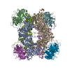
|
|---|---|
| 1 |
|
-Components
| #1: Protein | Mass: 72766.664 Da / Num. of mol.: 4 Source method: isolated from a genetically manipulated source Source: (gene. exp.) Homo sapiens (human) / Gene: PRMT5, HRMT1L5, IBP72, JBP1, SKB1 / Production host:  Trichoplusia ni (cabbage looper) Trichoplusia ni (cabbage looper)References: UniProt: O14744, type II protein arginine methyltransferase #2: Protein | Mass: 36757.246 Da / Num. of mol.: 4 Source method: isolated from a genetically manipulated source Source: (gene. exp.) Homo sapiens (human) / Gene: WDR77, MEP50, WD45, HKMT1069, Nbla10071 / Production host: Trichoplusia ni (cabbage looper) / References: UniProt: Q9BQA1#3: Protein | Mass: 26038.957 Da / Num. of mol.: 4 Source method: isolated from a genetically manipulated source Source: (gene. exp.)  #4: Protein | Mass: 39572.793 Da / Num. of mol.: 4 Source method: isolated from a genetically manipulated source Source: (gene. exp.) Homo sapiens (human) / Gene: LSM11 / Production host: Trichoplusia ni (cabbage looper) / References: UniProt: P83369#5: Chemical | ChemComp-ADN /   Mass: 267.241 Da / Num. of mol.: 4 / Source method: obtained synthetically / Formula: C10H13N5O4 / Feature type: SUBJECT OF INVESTIGATION Mass: 267.241 Da / Num. of mol.: 4 / Source method: obtained synthetically / Formula: C10H13N5O4 / Feature type: SUBJECT OF INVESTIGATIONHas ligand of interest | Y | Has protein modification | N | |
|---|
-Experimental details
-Experiment
| Experiment | Method: ELECTRON MICROSCOPY |
|---|---|
| EM experiment | Aggregation state: PARTICLE / 3D reconstruction method: single particle reconstruction |
- Sample preparation
| Component | Name: Methylosome / Type: COMPLEX / Entity ID: #1-#4 / Source: RECOMBINANT |
|---|---|
| Molecular weight | Experimental value: NO |
| Source (natural) | Organism: Homo sapiens (human) |
| Source (recombinant) | Organism: Trichoplusia ni (cabbage looper) |
| Buffer solution | pH: 8 |
| Specimen | Embedding applied: NO / Shadowing applied: NO / Staining applied: NO / Vitrification applied: YES |
| Vitrification | Cryogen name: ETHANE |
- Electron microscopy imaging
| Experimental equipment |  Model: Titan Krios / Image courtesy: FEI Company |
|---|---|
| Microscopy | Model: FEI TITAN KRIOS |
| Electron gun | Electron source:  FIELD EMISSION GUN / Accelerating voltage: 300 kV / Illumination mode: FLOOD BEAM FIELD EMISSION GUN / Accelerating voltage: 300 kV / Illumination mode: FLOOD BEAM |
| Electron lens | Mode: BRIGHT FIELD / Nominal defocus max: 1250 nm / Nominal defocus min: 800 nm |
| Image recording | Electron dose: 58.2 e/Å2 / Film or detector model: GATAN K3 (6k x 4k) |
- Processing
| Software | Name: PHENIX / Version: 1.18.2_3874: / Classification: refinement | ||||||||||||||||||||||||
|---|---|---|---|---|---|---|---|---|---|---|---|---|---|---|---|---|---|---|---|---|---|---|---|---|---|
| EM software | Name: PHENIX / Category: model refinement | ||||||||||||||||||||||||
| CTF correction | Type: PHASE FLIPPING AND AMPLITUDE CORRECTION | ||||||||||||||||||||||||
| 3D reconstruction | Resolution: 2.83 Å / Resolution method: FSC 0.143 CUT-OFF / Num. of particles: 754047 / Symmetry type: POINT | ||||||||||||||||||||||||
| Refine LS restraints |
|
