[English] 日本語
Yorodumi
- PDB-8fwh: Crystal structure of bivalent antibody Fab fragment of Anti-human... -

+
Open data


ID or keywords:

Loading...

-
Basic information

Entry
Database: PDB / ID: 8fwh
TitleCrystal structure of bivalent antibody Fab fragment of Anti-human LAG3 (22D2)
Components
  • Anti-human LAG3 (22D2) heavy chain
  • Anti-human LAG3 (22D2) light chain
KeywordsIMMUNE SYSTEM / Fab / inhibitory receptors
Biological speciesMus musculus (house mouse)
MethodX-RAY DIFFRACTION / SYNCHROTRON / MOLECULAR REPLACEMENT / Resolution: 2.833 Å
AuthorsMishra, A.K. / Agnihotri, P. / Mariuzza, R.A.
Funding support United States, 1items
OrganizationGrant numberCountry
National Institutes of Health/National Institute Of Allergy and Infectious Diseases (NIH/NIAID)AI144422 United States
Citation
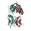
Journal: Structure / Year: 2023
Title: CryoEM structure of a therapeutic antibody (favezelimab) bound to human LAG3 determined using a bivalent Fab as fiducial marker.
Authors: Arjun K Mishra / Salman Shahid / Sharanbasappa S Karade / Pragati Agnihotri / Alexander Kolesnikov / S Saif Hasan / Roy A Mariuzza /
Abstract: Lymphocyte activation gene 3 protein (LAG3) is an inhibitory receptor that is upregulated on exhausted T cells in tumors. LAG3 is a major target for cancer immunotherapy with many anti-LAG3 ...Lymphocyte activation gene 3 protein (LAG3) is an inhibitory receptor that is upregulated on exhausted T cells in tumors. LAG3 is a major target for cancer immunotherapy with many anti-LAG3 antibodies in clinical trials. However, there is no structural information on the epitopes recognized by these antibodies. We determined the single-particle cryoEM structure of a therapeutic antibody (favezelimab) bound to LAG3 to 3.5 Å resolution, revealing that favezelimab targets the LAG3-binding site for MHC class II, its canonical ligand. The small size of the complex between the conventional (monovalent) Fab of favezelimab and LAG3 (∼100 kDa) presented a challenge for cryoEM. Accordingly, we engineered a bivalent version of Fab favezelimab that doubled the size of the Fab-LAG3 complex and conferred a highly identifiable shape to the complex that facilitated particle selection and orientation for image processing. This study establishes bivalent Fabs as new fiducial markers for cryoEM analysis of small proteins.
History
DepositionJan 22, 2023Deposition site: RCSB / Processing site: RCSB
Revision 1.0Sep 6, 2023Provider: repository / Type: Initial release
Revision 1.1Oct 18, 2023Group: Database references / Category: citation / Item: _citation.journal_volume / _citation.page_first

-
Structure visualization

Structure viewerMolecule:
MolmilJmol/JSmol

Downloads & links

-
Assembly

Deposited unit
HHH: Anti-human LAG3 (22D2) heavy chain
LLL: Anti-human LAG3 (22D2) light chain
hetero molecules


Theoretical massNumber of molelcules
Total (without water)47,7113
Polymers47,6492
Non-polymers621
Water48627
1
HHH: Anti-human LAG3 (22D2) heavy chain
LLL: Anti-human LAG3 (22D2) light chain
hetero molecules

HHH: Anti-human LAG3 (22D2) heavy chain
LLL: Anti-human LAG3 (22D2) light chain
hetero molecules


  • defined by author&software
  • Evidence: gel filtration
  • 95.4 kDa, 4 polymers
Theoretical massNumber of molelcules
Total (without water)95,4226
Polymers95,2984
Non-polymers1242
Water724
TypeNameSymmetry operationNumber
identity operation1_555x,y,z1
crystal symmetry operation2_555-x,y,-z1
Buried area6920 Å2
ΔGint-35 kcal/mol
Surface area38430 Å2
MethodPISA
Unit cell
Length a, b, c (Å)159.287, 44.390, 89.084
Angle α, β, γ (deg.)90.000, 106.136, 90.000
Int Tables number5
Space group name H-MC121

-
Components

#1: Antibody Anti-human LAG3 (22D2) heavy chain


Mass: 23959.947 Da / Num. of mol.: 1 / Fragment: Fab
Source method: isolated from a genetically manipulated source
Source: (gene. exp.) Mus musculus (house mouse) / Cell line (production host): HEK 293 expi / Production host: Homo sapiens (human)
#2: Antibody Anti-human LAG3 (22D2) light chain


Mass: 23688.984 Da / Num. of mol.: 1 / Fragment: Fab
Source method: isolated from a genetically manipulated source
Source: (gene. exp.) Mus musculus (house mouse) / Cell line (production host): HEK 293 expi / Production host: Homo sapiens (human)
#3: Chemical ChemComp-EDO / 1,2-ETHANEDIOL / ETHYLENE GLYCOL


Mass: 62.068 Da / Num. of mol.: 1 / Source method: obtained synthetically / Formula: C2H6O2 / Feature type: SUBJECT OF INVESTIGATION
#4: Water ChemComp-HOH / water


Mass: 18.015 Da / Num. of mol.: 27 / Source method: isolated from a natural source / Formula: H2O
Has ligand of interestY

-
Experimental details

-
Experiment

ExperimentMethod: X-RAY DIFFRACTION / Number of used crystals: 1

-
Sample preparation

CrystalDensity Matthews: 3.17 Å3/Da / Density % sol: 61.26 %
Crystal growTemperature: 295 K / Method: vapor diffusion, hanging drop / pH: 7.5
Details: 0.1 M Tris-HCl, pH 7.5, 0.2 M sodium chloride, 10% w/v PEG8000

-
Data collection

DiffractionMean temperature: 100 K / Serial crystal experiment: N
Diffraction sourceSource: SYNCHROTRON / Site: APS / Beamline: 23-ID-D / Wavelength: 1.03 Å
DetectorType: DECTRIS PILATUS 6M / Detector: PIXEL / Date: Mar 14, 2021
RadiationProtocol: SINGLE WAVELENGTH / Monochromatic (M) / Laue (L): M / Scattering type: x-ray
Radiation wavelengthWavelength: 1.03 Å / Relative weight: 1
ReflectionResolution: 2.83→85.57 Å / Num. obs: 13587 / % possible obs: 92.9 % / Redundancy: 2 % / CC1/2: 0.98 / Net I/σ(I): 13.7
Reflection shellResolution: 2.83→2.9 Å / Num. unique obs: 13515 / CC1/2: 0.68 / CC star: 0.9

-
Processing

Software
NameVersionClassification
REFMAC5.8.0267refinement
HKL-2000data reduction
HKL-2000data scaling
PHASERphasing
RefinementMethod to determine structure: MOLECULAR REPLACEMENT
Starting model: PDB entry 6WKM
Resolution: 2.833→42.75 Å / Cor.coef. Fo:Fc: 0.94 / Cor.coef. Fo:Fc free: 0.915 / SU B: 20.341 / SU ML: 0.365 / Cross valid method: FREE R-VALUE / ESU R: 2.956 / ESU R Free: 0.407
Details: Hydrogens have been added in their riding positions
RfactorNum. reflection% reflection
Rfree0.2767 706 5.196 %
Rwork0.2164 12881 -
all0.219 --
obs-13587 92.903 %
Solvent computationIon probe radii: 0.8 Å / Shrinkage radii: 0.8 Å / VDW probe radii: 1.2 Å / Solvent model: MASK BULK SOLVENT
Displacement parametersBiso mean: 77.32 Å2
Baniso -1Baniso -2Baniso -3
1--3.808 Å2-0 Å2-1.694 Å2
2--10.894 Å20 Å2
3----5.23 Å2
Refinement stepCycle: LAST / Resolution: 2.833→42.75 Å
ProteinNucleic acidLigandSolventTotal
Num. atoms3145 0 4 27 3176
Refine LS restraints
Refine-IDTypeDev idealDev ideal targetNumber
X-RAY DIFFRACTIONr_bond_refined_d0.0070.0133237
X-RAY DIFFRACTIONr_bond_other_d0.0010.0172846
X-RAY DIFFRACTIONr_angle_refined_deg1.3971.6434434
X-RAY DIFFRACTIONr_angle_other_deg1.2791.5696561
X-RAY DIFFRACTIONr_dihedral_angle_1_deg7.0195428
X-RAY DIFFRACTIONr_dihedral_angle_2_deg38.03923.459133
X-RAY DIFFRACTIONr_dihedral_angle_3_deg17.02415447
X-RAY DIFFRACTIONr_dihedral_angle_4_deg18.4741510
X-RAY DIFFRACTIONr_chiral_restr0.0520.2445
X-RAY DIFFRACTIONr_gen_planes_refined0.0050.023754
X-RAY DIFFRACTIONr_gen_planes_other0.0020.02722
X-RAY DIFFRACTIONr_nbd_refined0.2360.2503
X-RAY DIFFRACTIONr_symmetry_nbd_other0.2040.22698
X-RAY DIFFRACTIONr_nbtor_refined0.1630.21535
X-RAY DIFFRACTIONr_symmetry_nbtor_other0.0750.21666
X-RAY DIFFRACTIONr_xyhbond_nbd_refined0.1460.266
X-RAY DIFFRACTIONr_symmetry_nbd_refined0.1520.214
X-RAY DIFFRACTIONr_nbd_other0.1910.283
X-RAY DIFFRACTIONr_symmetry_xyhbond_nbd_refined0.2750.24
X-RAY DIFFRACTIONr_xyhbond_nbd_other0.270.21
X-RAY DIFFRACTIONr_mcbond_it6.0358.4171714
X-RAY DIFFRACTIONr_mcbond_other6.0348.4131712
X-RAY DIFFRACTIONr_mcangle_it9.18212.6192137
X-RAY DIFFRACTIONr_mcangle_other9.1812.6192137
X-RAY DIFFRACTIONr_scbond_it6.3328.7061523
X-RAY DIFFRACTIONr_scbond_other6.338.7051524
X-RAY DIFFRACTIONr_scangle_it9.76712.9112296
X-RAY DIFFRACTIONr_scangle_other9.76512.912297
X-RAY DIFFRACTIONr_lrange_it15.314160.14412936
X-RAY DIFFRACTIONr_lrange_other15.303160.14712934
LS refinement shell
Resolution (Å)Rfactor RfreeNum. reflection RfreeRfactor RworkNum. reflection RworkRefine-ID% reflection obs (%)
2.833-2.9070.467470.341754X-RAY DIFFRACTION73.2176
2.907-2.9860.342410.316841X-RAY DIFFRACTION86.3859
2.986-3.0730.363600.302875X-RAY DIFFRACTION91.4873
3.073-3.1670.37560.303871X-RAY DIFFRACTION93.8259
3.167-3.2710.376540.281853X-RAY DIFFRACTION96.0805
3.271-3.3850.276430.25840X-RAY DIFFRACTION96.1874
3.385-3.5130.311390.24805X-RAY DIFFRACTION95.2596
3.513-3.6560.284320.236785X-RAY DIFFRACTION95.1106
3.656-3.8180.291440.219749X-RAY DIFFRACTION96.0048
3.818-4.0040.241440.202726X-RAY DIFFRACTION96.01
4.004-4.220.196330.159677X-RAY DIFFRACTION96.206
4.22-4.4760.212390.162667X-RAY DIFFRACTION96.4481
4.476-4.7840.237400.169582X-RAY DIFFRACTION96.136
4.784-5.1660.262290.172591X-RAY DIFFRACTION95.679
5.166-5.6570.249280.171528X-RAY DIFFRACTION96.5278
5.657-6.3210.233210.208490X-RAY DIFFRACTION94.6296
6.321-7.2920.387240.234423X-RAY DIFFRACTION94.5032
7.292-8.9150.178120.185368X-RAY DIFFRACTION94.7631
8.915-12.540.222130.201282X-RAY DIFFRACTION94.2492
12.54-42.750.32970.319174X-RAY DIFFRACTION91.8782

+
About Yorodumi

-
News

-
Feb 9, 2022. New format data for meta-information of EMDB entries

New format data for meta-information of EMDB entries

  • Version 3 of the EMDB header file is now the official format.
  • The previous official version 1.9 will be removed from the archive.

Related info.:EMDB header

External links:wwPDB to switch to version 3 of the EMDB data model

-
Aug 12, 2020. Covid-19 info

Covid-19 info

URL: https://pdbjlvh1.pdbj.org/emnavi/covid19.php

New page: Covid-19 featured information page in EM Navigator.

Related info.:Covid-19 info / Mar 5, 2020. Novel coronavirus structure data

+
Mar 5, 2020. Novel coronavirus structure data

Novel coronavirus structure data

Related info.:Yorodumi Speices / Aug 12, 2020. Covid-19 info

External links:COVID-19 featured content - PDBj / Molecule of the Month (242):Coronavirus Proteases

+
Jan 31, 2019. EMDB accession codes are about to change! (news from PDBe EMDB page)

EMDB accession codes are about to change! (news from PDBe EMDB page)

  • The allocation of 4 digits for EMDB accession codes will soon come to an end. Whilst these codes will remain in use, new EMDB accession codes will include an additional digit and will expand incrementally as the available range of codes is exhausted. The current 4-digit format prefixed with “EMD-” (i.e. EMD-XXXX) will advance to a 5-digit format (i.e. EMD-XXXXX), and so on. It is currently estimated that the 4-digit codes will be depleted around Spring 2019, at which point the 5-digit format will come into force.
  • The EM Navigator/Yorodumi systems omit the EMD- prefix.

Related info.:Q: What is EMD? / ID/Accession-code notation in Yorodumi/EM Navigator

External links:EMDB Accession Codes are Changing Soon! / Contact to PDBj

+
Jul 12, 2017. Major update of PDB

Major update of PDB

  • wwPDB released updated PDB data conforming to the new PDBx/mmCIF dictionary.
  • This is a major update changing the version number from 4 to 5, and with Remediation, in which all the entries are updated.
  • In this update, many items about electron microscopy experimental information are reorganized (e.g. em_software).
  • Now, EM Navigator and Yorodumi are based on the updated data.

External links:wwPDB Remediation / Enriched Model Files Conforming to OneDep Data Standards Now Available in the PDB FTP Archive

-
Yorodumi

Thousand views of thousand structures

  • Yorodumi is a browser for structure data from EMDB, PDB, SASBDB, etc.
  • This page is also the successor to EM Navigator detail page, and also detail information page/front-end page for Omokage search.
  • The word "yorodu" (or yorozu) is an old Japanese word meaning "ten thousand". "mi" (miru) is to see.

Related info.:EMDB / PDB / SASBDB / Comparison of 3 databanks / Yorodumi Search / Aug 31, 2016. New EM Navigator & Yorodumi / Yorodumi Papers / Jmol/JSmol / Function and homology information / Changes in new EM Navigator and Yorodumi

Read more