 Movie
Movie Controller


+ Open data

- Basic information
| Entry | Database: PDB / ID: 8fvb | |||||||||
|---|---|---|---|---|---|---|---|---|---|---|
| Title | E coli. CTP synthase in complex with CTP | |||||||||
 Components Components | CTP synthase | |||||||||
 Keywords Keywords | CYTOSOLIC PROTEIN / LIGASE / Inhibitor complex / Metabolic enzyme | |||||||||
| Function / homology |  Function and homology information Function and homology informationCTP synthase (glutamine hydrolysing) / CTP synthase activity / cytoophidium / 'de novo' CTP biosynthetic process / pyrimidine nucleobase biosynthetic process / glutaminase activity / ATP binding / metal ion binding / identical protein binding / cytosol Similarity search - Function | |||||||||
| Biological species |  | |||||||||
| Method |  X-RAY DIFFRACTION / SYNCHROTRON / MOLECULAR REPLACEMENT / Resolution: 2.03 Å X-RAY DIFFRACTION / SYNCHROTRON / MOLECULAR REPLACEMENT / Resolution: 2.03 Å | |||||||||
 Authors Authors | Holyoak, T. / McLeod, M.J. / Tran, N. | |||||||||
| Funding support |  Canada, 2items Canada, 2items
| |||||||||
 Citation Citation | Journal: Protein Sci. / Year: 2023 Title: A metal-dependent conformational change provides a structural basis for the inhibition of CTP synthase by gemcitabine-5'-triphosphate. Authors: McLeod, M.J. / Tran, N. / McCluskey, G.D. / Gillis, T.D. / Bearne, S.L. / Holyoak, T. | |||||||||
| History |
|
- Structure visualization
| Structure viewer | Molecule: MolmilJmol/JSmol |
|---|
- Downloads & links
-Download
| PDBx/mmCIF format | 8fvb.cif.gz | 252.9 KB | Display | PDBx/mmCIF format |
|---|---|---|---|---|
| PDB format | pdb8fvb.ent.gz | Display | PDB format | |
| PDBx/mmJSON format | 8fvb.json.gz | Tree view | PDBx/mmJSON format | |
| Others |  Other downloads Other downloads |
-Validation report
| Arichive directory | https://data.pdbj.org/pub/pdb/validation_reports/fv/8fvbftp://data.pdbj.org/pub/pdb/validation_reports/fv/8fvb | HTTPS FTP |
|---|
-Related structure data
| Related structure data | 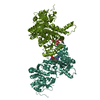 8fv6C  8fv7C 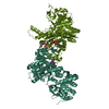 8fv8C 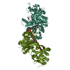 8fv9C 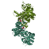 8fvaC  8fvcC  8fvdC  8fveC  8sbrC 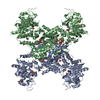 5tkvS S: Starting model for refinement C: citing same article ( |
|---|---|
| Similar structure data |
-Links
 PDBj
PDBj


- Assembly
| Deposited unit | 
| ||||||||
|---|---|---|---|---|---|---|---|---|---|
| 1 | 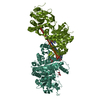
| ||||||||
| Unit cell |
| ||||||||
| Components on special symmetry positions |
|
-Components
| #1: Protein | Mass: 60446.980 Da / Num. of mol.: 2 Source method: isolated from a genetically manipulated source Source: (gene. exp.) References: UniProt: B7MLA1, CTP synthase (glutamine hydrolysing) #2: Chemical | ChemComp-MLA /   Mass: 104.061 Da / Num. of mol.: 4 / Source method: obtained synthetically / Formula: C3H4O4 Mass: 104.061 Da / Num. of mol.: 4 / Source method: obtained synthetically / Formula: C3H4O4#3: Chemical |   Mass: 483.156 Da / Num. of mol.: 2 / Source method: obtained synthetically / Formula: C9H16N3O14P3 / Feature type: SUBJECT OF INVESTIGATION Mass: 483.156 Da / Num. of mol.: 2 / Source method: obtained synthetically / Formula: C9H16N3O14P3 / Feature type: SUBJECT OF INVESTIGATION#4: Water | ChemComp-HOH / |  Mass: 18.015 Da / Num. of mol.: 847 / Source method: isolated from a natural source / Formula: H2O Mass: 18.015 Da / Num. of mol.: 847 / Source method: isolated from a natural source / Formula: H2OHas ligand of interest | Y | |
|---|
-Experimental details
-Experiment
| Experiment | Method: X-RAY DIFFRACTION / Number of used crystals: 1 |
|---|
- Sample preparation
| Crystal | Density Matthews: 4.54 Å3/Da / Density % sol: 72.94 % |
|---|---|
| Crystal grow | Temperature: 277 K / Method: vapor diffusion, hanging drop / pH: 8 Details: 0.1 M TRIS-Cl pH 8.0 (room temperature), 5 mM magnesium chloride, 1.15 - 1.4 M ammonium sulfate, 15 mg/mL CTPS. |
-Data collection
| Diffraction | Mean temperature: 100 K / Serial crystal experiment: N |
|---|---|
| Diffraction source | Source: SYNCHROTRON / Site: CLSI / Beamline: 08B1-1 / Wavelength: 0.978 Å |
| Detector | Type: RAYONIX MX-300 / Detector: CCD / Date: May 9, 2019 |
| Radiation | Protocol: SINGLE WAVELENGTH / Monochromatic (M) / Laue (L): M / Scattering type: x-ray |
| Radiation wavelength | Wavelength: 0.978 Å / Relative weight: 1 |
| Reflection | Resolution: 2.03→44 Å / Num. obs: 142127 / % possible obs: 97.3 % / Redundancy: 14 % / Biso Wilson estimate: 33.4 Å2 / CC1/2: 0.97 / CC star: 0.992 / Rmerge(I) obs: 0.166 / Rpim(I) all: 0.045 / Rrim(I) all: 0.172 / Net I/σ(I): 4.83 |
| Reflection shell | Resolution: 2.03→2.1 Å / Redundancy: 12.1 % / Rmerge(I) obs: 0.851 / Mean I/σ(I) obs: 1.15 / Num. unique obs: 10569 / CC1/2: 0.892 / CC star: 0.971 / Rpim(I) all: 0.248 / Rrim(I) all: 0.887 / % possible all: 73.4 |
- Processing
| Software |
| ||||||||||||||||||||||||||||||||||||||||||||||||||||||||||||||||||||||||||||||||||||||||||||||||||||||||||||||||||||||||||||||||||||||||||||||||||||||
|---|---|---|---|---|---|---|---|---|---|---|---|---|---|---|---|---|---|---|---|---|---|---|---|---|---|---|---|---|---|---|---|---|---|---|---|---|---|---|---|---|---|---|---|---|---|---|---|---|---|---|---|---|---|---|---|---|---|---|---|---|---|---|---|---|---|---|---|---|---|---|---|---|---|---|---|---|---|---|---|---|---|---|---|---|---|---|---|---|---|---|---|---|---|---|---|---|---|---|---|---|---|---|---|---|---|---|---|---|---|---|---|---|---|---|---|---|---|---|---|---|---|---|---|---|---|---|---|---|---|---|---|---|---|---|---|---|---|---|---|---|---|---|---|---|---|---|---|---|---|---|---|
| Refinement | Method to determine structure: MOLECULAR REPLACEMENT Starting model: 5TKV Resolution: 2.03→44 Å / Cor.coef. Fo:Fc: 0.957 / Cor.coef. Fo:Fc free: 0.946 / SU B: 3.164 / SU ML: 0.086 / Cross valid method: FREE R-VALUE / ESU R: 0.125 / ESU R Free: 0.118 Details: Hydrogens have been added in their riding positions
| ||||||||||||||||||||||||||||||||||||||||||||||||||||||||||||||||||||||||||||||||||||||||||||||||||||||||||||||||||||||||||||||||||||||||||||||||||||||
| Solvent computation | Ion probe radii: 0.8 Å / Shrinkage radii: 0.8 Å / VDW probe radii: 1.2 Å / Solvent model: MASK BULK SOLVENT | ||||||||||||||||||||||||||||||||||||||||||||||||||||||||||||||||||||||||||||||||||||||||||||||||||||||||||||||||||||||||||||||||||||||||||||||||||||||
| Displacement parameters | Biso mean: 35.822 Å2
| ||||||||||||||||||||||||||||||||||||||||||||||||||||||||||||||||||||||||||||||||||||||||||||||||||||||||||||||||||||||||||||||||||||||||||||||||||||||
| Refinement step | Cycle: LAST / Resolution: 2.03→44 Å
| ||||||||||||||||||||||||||||||||||||||||||||||||||||||||||||||||||||||||||||||||||||||||||||||||||||||||||||||||||||||||||||||||||||||||||||||||||||||
| Refine LS restraints |
| ||||||||||||||||||||||||||||||||||||||||||||||||||||||||||||||||||||||||||||||||||||||||||||||||||||||||||||||||||||||||||||||||||||||||||||||||||||||
| LS refinement shell |
|
