 Movie
Movie Controller

[English] 日本語
 Yorodumi
- PDB-8fv1: SpeG spermidine N-acetyltransferase from Staphylococcus aureus in... -
+ Open data

- Basic information
| Entry | Database: PDB / ID: 8fv1 | |||||||||
|---|---|---|---|---|---|---|---|---|---|---|
| Title | SpeG spermidine N-acetyltransferase from Staphylococcus aureus in complex with spermine | |||||||||
 Components Components | Diamine N-acetyltransferase | |||||||||
 Keywords Keywords | TRANSFERASE | |||||||||
| Function / homology | diamine N-acetyltransferase activity / Acetyltransferase (GNAT) domain / Gcn5-related N-acetyltransferase (GNAT) domain profile. / GNAT domain / Acyl-CoA N-acyltransferase / SPERMINE / Diamine N-acetyltransferase Function and homology information Function and homology information | |||||||||
| Biological species |   Staphylococcus aureus (bacteria) Staphylococcus aureus (bacteria) | |||||||||
| Method |  X-RAY DIFFRACTION / SYNCHROTRON / MOLECULAR REPLACEMENT / Resolution: 2.95 Å X-RAY DIFFRACTION / SYNCHROTRON / MOLECULAR REPLACEMENT / Resolution: 2.95 Å | |||||||||
 Authors Authors | Tsimbalyuk, S. / Forwood, J.K. | |||||||||
| Funding support | 1items
| |||||||||
 Citation Citation | Journal: To Be Published Title: SpeG spermidine N-acetyltransferase from Staphylococcus aureus in complex with spermine, crystal form II Authors: Tsimbalyuk, S. / Forwood, J.K. | |||||||||
| History |
|
- Structure visualization
| Structure viewer | Molecule: MolmilJmol/JSmol |
|---|
- Downloads & links
-Download
| PDBx/mmCIF format | 8fv1.cif.gz | 326.8 KB | Display | PDBx/mmCIF format |
|---|---|---|---|---|
| PDB format | pdb8fv1.ent.gz | 219.3 KB | Display | PDB format |
| PDBx/mmJSON format | 8fv1.json.gz | Tree view | PDBx/mmJSON format | |
| Others |  Other downloads Other downloads |
-Validation report
| Arichive directory | https://data.pdbj.org/pub/pdb/validation_reports/fv/8fv1ftp://data.pdbj.org/pub/pdb/validation_reports/fv/8fv1 | HTTPS FTP |
|---|
-Related structure data
| Related structure data | 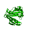 5ix3S S: Starting model for refinement |
|---|---|
| Similar structure data |
-Links
 PDBj
PDBj


- Assembly
| Deposited unit | 
| ||||||||||||
|---|---|---|---|---|---|---|---|---|---|---|---|---|---|
| 1 | 
| ||||||||||||
| Unit cell |
|
-Components
| #1: Protein | Mass: 20077.939 Da / Num. of mol.: 4 Source method: isolated from a genetically manipulated source Source: (gene. exp.) Staphylococcus aureus (bacteria) / Gene: res / Production host: #2: Chemical | ChemComp-SPM /   Mass: 202.340 Da / Num. of mol.: 4 / Source method: obtained synthetically / Formula: C10H26N4 Mass: 202.340 Da / Num. of mol.: 4 / Source method: obtained synthetically / Formula: C10H26N4Has ligand of interest | N | |
|---|
-Experimental details
-Experiment
| Experiment | Method: X-RAY DIFFRACTION / Number of used crystals: 1 |
|---|
- Sample preparation
| Crystal | Density Matthews: 4.36 Å3/Da / Density % sol: 71.77 % |
|---|---|
| Crystal grow | Temperature: 290 K / Method: vapor diffusion, hanging drop / Details: 0.1M NaCl, 0.1M HEPES pH8.0, 1.6M Ammonium Sulfate |
-Data collection
| Diffraction | Mean temperature: 100 K / Serial crystal experiment: N |
|---|---|
| Diffraction source | Source: SYNCHROTRON / Site: Australian Synchrotron  / Beamline: MX2 / Wavelength: 0.9762 Å / Beamline: MX2 / Wavelength: 0.9762 Å |
| Detector | Type: ADSC QUANTUM 315r / Detector: CCD / Date: Jul 10, 2020 |
| Radiation | Protocol: SINGLE WAVELENGTH / Monochromatic (M) / Laue (L): M / Scattering type: x-ray |
| Radiation wavelength | Wavelength: 0.9762 Å / Relative weight: 1 |
| Reflection | Resolution: 2.95→24.95 Å / Num. obs: 29846 / % possible obs: 99.75 % / Redundancy: 20 % / Biso Wilson estimate: 77.58 Å2 / CC1/2: 0.999 / Net I/σ(I): 18.9 |
| Reflection shell | Resolution: 2.95→3.13 Å / Num. unique obs: 2792 / CC1/2: 0.681 |
- Processing
| Software |
| |||||||||||||||||||||||||||||||||||||||||||||||||||||||||||||||||||||||||||||
|---|---|---|---|---|---|---|---|---|---|---|---|---|---|---|---|---|---|---|---|---|---|---|---|---|---|---|---|---|---|---|---|---|---|---|---|---|---|---|---|---|---|---|---|---|---|---|---|---|---|---|---|---|---|---|---|---|---|---|---|---|---|---|---|---|---|---|---|---|---|---|---|---|---|---|---|---|---|---|
| Refinement | Method to determine structure: MOLECULAR REPLACEMENT Starting model: 5ix3 Resolution: 2.95→24.95 Å / SU ML: 0.4095 / Cross valid method: FREE R-VALUE / σ(F): 1.34 / Phase error: 29.9408 Stereochemistry target values: GeoStd + Monomer Library + CDL v1.2
| |||||||||||||||||||||||||||||||||||||||||||||||||||||||||||||||||||||||||||||
| Solvent computation | Shrinkage radii: 0.9 Å / VDW probe radii: 1.1 Å / Solvent model: FLAT BULK SOLVENT MODEL | |||||||||||||||||||||||||||||||||||||||||||||||||||||||||||||||||||||||||||||
| Displacement parameters | Biso mean: 80.41 Å2 | |||||||||||||||||||||||||||||||||||||||||||||||||||||||||||||||||||||||||||||
| Refinement step | Cycle: LAST / Resolution: 2.95→24.95 Å
| |||||||||||||||||||||||||||||||||||||||||||||||||||||||||||||||||||||||||||||
| Refine LS restraints |
| |||||||||||||||||||||||||||||||||||||||||||||||||||||||||||||||||||||||||||||
| LS refinement shell |
|
