| Entry | Database: PDB / ID: 8fut
|
|---|
| Title | Crystal structure of Xenopus laevis arrestin 1 - P3121 crystal form |
|---|
 Components Components | S-arrestin |
|---|
 Keywords Keywords | SIGNALING PROTEIN / arrestin 1 / visual arrestin / S-antigen |
|---|
| Function / homology |  Function and homology information Function and homology information
G protein-coupled receptor internalization / photoreceptor outer segment / photoreceptor inner segment / visual perception / G protein-coupled receptor binding / signal transduction / membraneSimilarity search - Function Arrestin, conserved site / Arrestins signature. / Arrestin / Arrestin, N-terminal / Arrestin-like, N-terminal / Arrestin C-terminal-like domain / Arrestin (or S-antigen), N-terminal domain / Arrestin (or S-antigen), C-terminal domain / Arrestin (or S-antigen), C-terminal domain / Arrestin-like, C-terminal / Immunoglobulin E-setSimilarity search - Domain/homology |
|---|
| Biological species |  Xenopus laevis (African clawed frog) Xenopus laevis (African clawed frog) |
|---|
| Method |  X-RAY DIFFRACTION / SYNCHROTRON / FOURIER SYNTHESIS / Resolution: 2.54 Å X-RAY DIFFRACTION / SYNCHROTRON / FOURIER SYNTHESIS / Resolution: 2.54 Å |
|---|
 Authors Authors | Salom, D. / Barnes, C.L. / Calvert, P.D. / Kiser, P.D. |
|---|
| Funding support |  United States, 3items United States, 3items | Organization | Grant number | Country |
|---|
| National Institutes of Health/National Eye Institute (NIH/NEI) | R01EY018421 | United States | | National Institutes of Health/National Eye Institute (NIH/NEI) | R01EY028303 | United States | | Department of Veterans Affairs (VA, United States) | I01 BX004939 | United States |
|
|---|
 Citation Citation | Journal: J.Biol.Chem. / Year: 2024
Title: Mechanisms of amphibian arrestin 1 self-association and dynamic distribution in retinal photoreceptors.
Authors: Barnes, C.L. / Salom, D. / Namitz, K.E.W. / Smith, W.C. / Knutson, B.A. / Cosgrove, M.S. / Kiser, P.D. / Calvert, P.D. |
|---|
| History | | Deposition | Jan 18, 2023 | Deposition site: RCSB / Processing site: RCSB |
|---|
| Revision 1.0 | Jul 24, 2024 | Provider: repository / Type: Initial release |
|---|
| Revision 1.1 | Dec 18, 2024 | Group: Database references / Structure summary / Category: citation / citation_author / pdbx_entry_details
Item: _citation.country / _citation.journal_abbrev ..._citation.country / _citation.journal_abbrev / _citation.journal_id_ASTM / _citation.journal_id_CSD / _citation.journal_id_ISSN / _citation.journal_volume / _citation.page_first / _citation.page_last / _citation.pdbx_database_id_DOI / _citation.pdbx_database_id_PubMed / _citation.title / _citation.year |
|---|
|
|---|
 Movie
Movie Controller

 Yorodumi
 Open data
 Basic information
 Structure visualization
 Downloads & links
 Other downloads
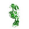
 PDBj
PDBj Assembly
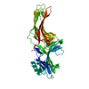

 Mass: 18.015 Da / Num. of mol.: 22 / Source method: isolated from a natural source / Formula: H2O
 Sample preparation
 Processing