 Movie
Movie Controller

[English] 日本語
 Yorodumi
- PDB-8fub: Crystal structure of human Importin alpha 3 in complex with Hendr... -
+ Open data

- Basic information
| Entry | Database: PDB / ID: 8fub | ||||||
|---|---|---|---|---|---|---|---|
| Title | Crystal structure of human Importin alpha 3 in complex with Hendra virus matrix protein NLS1 | ||||||
 Components Components |
| ||||||
 Keywords Keywords | TRANSPORT PROTEIN / Importin / nuclear transport | ||||||
| Function / homology |  Function and homology information Function and homology informationdopamine secretion / NS1 Mediated Effects on Host Pathways / NLS-dependent protein nuclear import complex / NLS-bearing protein import into nucleus / nuclear localization sequence binding / nuclear import signal receptor activity / virion assembly / nuclear pore / male germ cell nucleus / response to hydrogen peroxide ...dopamine secretion / NS1 Mediated Effects on Host Pathways / NLS-dependent protein nuclear import complex / NLS-bearing protein import into nucleus / nuclear localization sequence binding / nuclear import signal receptor activity / virion assembly / nuclear pore / male germ cell nucleus / response to hydrogen peroxide / virion component / ISG15 antiviral mechanism / protein import into nucleus / nuclear membrane / gene expression / structural constituent of virion / host cell cytoplasm / host cell nucleus / nucleoplasm / nucleus / cytosol / cytoplasm Similarity search - Function | ||||||
| Biological species |  Homo sapiens (human) Homo sapiens (human) Hendra virus horse/Australia/Hendra/1994 Hendra virus horse/Australia/Hendra/1994 | ||||||
| Method |  X-RAY DIFFRACTION / SYNCHROTRON / MOLECULAR REPLACEMENT / Resolution: 2.75 Å X-RAY DIFFRACTION / SYNCHROTRON / MOLECULAR REPLACEMENT / Resolution: 2.75 Å | ||||||
 Authors Authors | Donnelly, C.M. / Basler, C.F. / Scott, C. / Forwood, J.K. | ||||||
| Funding support |  United States, 1items United States, 1items
| ||||||
 Citation Citation | Journal: Viruses / Year: 2023 Title: Henipavirus Matrix Protein Employs a Non-Classical Nuclear Localization Signal Binding Mechanism. Authors: Donnelly, C.M. / Vogel, O.A. / Edwards, M.R. / Taylor, P.E. / Roby, J.A. / Forwood, J.K. / Basler, C.F. | ||||||
| History |
|
- Structure visualization
| Structure viewer | Molecule: MolmilJmol/JSmol |
|---|
- Downloads & links
-Download
| PDBx/mmCIF format | 8fub.cif.gz | 114 KB | Display | PDBx/mmCIF format |
|---|---|---|---|---|
| PDB format | pdb8fub.ent.gz | 69.1 KB | Display | PDB format |
| PDBx/mmJSON format | 8fub.json.gz | Tree view | PDBx/mmJSON format | |
| Others |  Other downloads Other downloads |
-Validation report
| Summary document | 8fub_validation.pdf.gz | 430.9 KB | Display | wwPDB validaton report |
|---|---|---|---|---|
| Full document | 8fub_full_validation.pdf.gz | 435.2 KB | Display | |
| Data in XML | 8fub_validation.xml.gz | 15.7 KB | Display | |
| Data in CIF | 8fub_validation.cif.gz | 20.6 KB | Display | |
| Arichive directory | https://data.pdbj.org/pub/pdb/validation_reports/fu/8fubftp://data.pdbj.org/pub/pdb/validation_reports/fu/8fub | HTTPS FTP |
-Related structure data
| Related structure data |  8fuaC  8fucC  6bvzS S: Starting model for refinement C: citing same article ( |
|---|---|
| Similar structure data |
-Links
 PDBj
PDBj


- Assembly
| Deposited unit | 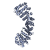
| ||||||||||||
|---|---|---|---|---|---|---|---|---|---|---|---|---|---|
| 1 |
| ||||||||||||
| Unit cell |
|
-Components
| #1: Protein | Mass: 50325.812 Da / Num. of mol.: 1 Source method: isolated from a genetically manipulated source Source: (gene. exp.) Homo sapiens (human) / Gene: KPNA4, QIP1 / Production host:  |
|---|---|
| #2: Protein/peptide | Mass: 2136.585 Da / Num. of mol.: 1 / Source method: obtained synthetically / Source: (synth.) Hendra virus horse/Australia/Hendra/1994 / References: UniProt: O89341 |
-Experimental details
-Experiment
| Experiment | Method: X-RAY DIFFRACTION / Number of used crystals: 1 |
|---|
- Sample preparation
| Crystal | Density Matthews: 2.5 Å3/Da / Density % sol: 50.83 % |
|---|---|
| Crystal grow | Temperature: 293 K / Method: vapor diffusion, hanging drop / pH: 7 Details: 0.1M sodium HEPES,0.72M sodium citrate, 1% dithiothreitol |
-Data collection
| Diffraction | Mean temperature: 100 K / Serial crystal experiment: N |
|---|---|
| Diffraction source | Source: SYNCHROTRON / Site: Australian Synchrotron  / Beamline: MX2 / Wavelength: 0.9537 Å / Beamline: MX2 / Wavelength: 0.9537 Å |
| Detector | Type: DECTRIS EIGER2 S 16M / Detector: PIXEL / Date: Jul 25, 2019 |
| Radiation | Monochromator: M / Protocol: SINGLE WAVELENGTH / Monochromatic (M) / Laue (L): M / Scattering type: x-ray |
| Radiation wavelength | Wavelength: 0.9537 Å / Relative weight: 1 |
| Reflection | Resolution: 2.75→29.78 Å / Num. obs: 13681 / % possible obs: 99.9 % / Redundancy: 11.3 % / Biso Wilson estimate: 65.77 Å2 / CC1/2: 0.995 / Rmerge(I) obs: 0.181 / Net I/σ(I): 7.8 |
| Reflection shell | Resolution: 2.75→2.9 Å / Rmerge(I) obs: 1.032 / Mean I/σ(I) obs: 2.3 / Num. unique obs: 1966 / CC1/2: 0.887 / Rpim(I) all: 0.446 |
- Processing
| Software |
| ||||||||||||||||||||||||||||||||||||||||||
|---|---|---|---|---|---|---|---|---|---|---|---|---|---|---|---|---|---|---|---|---|---|---|---|---|---|---|---|---|---|---|---|---|---|---|---|---|---|---|---|---|---|---|---|
| Refinement | Method to determine structure: MOLECULAR REPLACEMENT Starting model: 6BVZ Resolution: 2.75→29.78 Å / SU ML: 0.3291 / Cross valid method: FREE R-VALUE / σ(F): 1.34 / Phase error: 30.2044 / Stereochemistry target values: CDL v1.2
| ||||||||||||||||||||||||||||||||||||||||||
| Solvent computation | Shrinkage radii: 0.9 Å / VDW probe radii: 1.11 Å / Solvent model: FLAT BULK SOLVENT MODEL | ||||||||||||||||||||||||||||||||||||||||||
| Displacement parameters | Biso mean: 72.53 Å2 | ||||||||||||||||||||||||||||||||||||||||||
| Refinement step | Cycle: LAST / Resolution: 2.75→29.78 Å
| ||||||||||||||||||||||||||||||||||||||||||
| Refine LS restraints |
| ||||||||||||||||||||||||||||||||||||||||||
| LS refinement shell |
|
