 Movie
Movie Controller

[English] 日本語
 Yorodumi
- PDB-8ftg: Biophysical and Structural Characterization of an Anti-Caffeine V... -
+ Open data

- Basic information
| Entry | Database: PDB / ID: 8ftg | ||||||
|---|---|---|---|---|---|---|---|
| Title | Biophysical and Structural Characterization of an Anti-Caffeine VHH Antibody | ||||||
 Components Components | Anti-Caffeine VHH Antibody | ||||||
 Keywords Keywords | IMMUNE SYSTEM / camelid antibody / nanobody / hapten recognition / VHH / dimerization / chemically-induced dimerization (CID) system | ||||||
| Function / homology | Immunoglobulins / Immunoglobulin-like / Sandwich / Mainly Beta / CAFFEINE Function and homology information Function and homology information | ||||||
| Biological species |  | ||||||
| Method |  X-RAY DIFFRACTION / SYNCHROTRON / MOLECULAR REPLACEMENT / Resolution: 1.13 Å X-RAY DIFFRACTION / SYNCHROTRON / MOLECULAR REPLACEMENT / Resolution: 1.13 Å | ||||||
 Authors Authors | Horn, J.R. / Smith, C.A. / Sonneson, G.J. / Walter, R. | ||||||
| Funding support |  United States, 1items United States, 1items
| ||||||
 Citation Citation | Journal: Mabs / Year: 2023 Title: Molecular recognition requires dimerization of a VHH antibody. Authors: Smith, C.A. / Sonneson, G.J. / Hoey, R.J. / Hinerman, J.M. / Sheehy, K. / Walter, R. / Herr, A.B. / Horn, J.R. | ||||||
| History |
|
- Structure visualization
| Structure viewer | Molecule: MolmilJmol/JSmol |
|---|
- Downloads & links
-Download
| PDBx/mmCIF format | 8ftg.cif.gz | 663.3 KB | Display | PDBx/mmCIF format |
|---|---|---|---|---|
| PDB format | pdb8ftg.ent.gz | 458.6 KB | Display | PDB format |
| PDBx/mmJSON format | 8ftg.json.gz | Tree view | PDBx/mmJSON format | |
| Others |  Other downloads Other downloads |
-Validation report
| Arichive directory | https://data.pdbj.org/pub/pdb/validation_reports/ft/8ftgftp://data.pdbj.org/pub/pdb/validation_reports/ft/8ftg | HTTPS FTP |
|---|
-Related structure data
| Similar structure data |
|---|
-Links
 PDBj
PDBj


- Assembly
| Deposited unit | 
| ||||||||||||
|---|---|---|---|---|---|---|---|---|---|---|---|---|---|
| 1 | 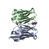
| ||||||||||||
| 2 | 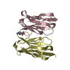
| ||||||||||||
| 3 | 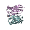
| ||||||||||||
| 4 | 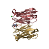
| ||||||||||||
| Unit cell |
|
-Components
| #1: Antibody | Mass: 13166.660 Da / Num. of mol.: 8 Source method: isolated from a genetically manipulated source Details: CDR graft into anti-RNaseA VHH Framework / Source: (gene. exp.)  #2: Chemical | ChemComp-CFF / 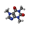  Mass: 194.191 Da / Num. of mol.: 4 / Source method: obtained synthetically / Formula: C8H10N4O2 / Feature type: SUBJECT OF INVESTIGATION / Comment: medication*YM Mass: 194.191 Da / Num. of mol.: 4 / Source method: obtained synthetically / Formula: C8H10N4O2 / Feature type: SUBJECT OF INVESTIGATION / Comment: medication*YM#3: Chemical | ChemComp-TRS / |   Mass: 122.143 Da / Num. of mol.: 1 / Source method: obtained synthetically / Formula: C4H12NO3 / Comment: pH buffer*YM Mass: 122.143 Da / Num. of mol.: 1 / Source method: obtained synthetically / Formula: C4H12NO3 / Comment: pH buffer*YM#4: Chemical |   Mass: 35.453 Da / Num. of mol.: 3 / Source method: obtained synthetically / Formula: Cl Mass: 35.453 Da / Num. of mol.: 3 / Source method: obtained synthetically / Formula: Cl#5: Water | ChemComp-HOH / |  Mass: 18.015 Da / Num. of mol.: 950 / Source method: isolated from a natural source / Formula: H2O Mass: 18.015 Da / Num. of mol.: 950 / Source method: isolated from a natural source / Formula: H2OHas ligand of interest | Y | |
|---|
-Experimental details
-Experiment
| Experiment | Method: X-RAY DIFFRACTION / Number of used crystals: 1 |
|---|
- Sample preparation
| Crystal | Density Matthews: 2.01 Å3/Da / Density % sol: 38.92 % |
|---|---|
| Crystal grow | Temperature: 293 K / Method: vapor diffusion, hanging drop / pH: 5.5 Details: The VHH/caffeine complex was crystallized using the hanging-drop vapor diffusion method. The drops contained 2 microliters of the 20 mg/ml complex and 2 microliters of the well mother liquor ...Details: The VHH/caffeine complex was crystallized using the hanging-drop vapor diffusion method. The drops contained 2 microliters of the 20 mg/ml complex and 2 microliters of the well mother liquor which contained 0.1 M BIS-TRIS (pH 5.5) and 25% w/v polyethylene glycol 3,350. |
-Data collection
| Diffraction | Mean temperature: 100 K / Serial crystal experiment: N |
|---|---|
| Diffraction source | Source: SYNCHROTRON / Site: APS / Beamline: 22-BM / Wavelength: 1 Å |
| Detector | Type: MAR CCD 165 mm / Detector: CCD / Date: Dec 1, 2008 |
| Radiation | Protocol: SINGLE WAVELENGTH / Monochromatic (M) / Laue (L): M / Scattering type: x-ray |
| Radiation wavelength | Wavelength: 1 Å / Relative weight: 1 |
| Reflection | Resolution: 1.13→50 Å / Num. obs: 287785 / % possible obs: 93.81 % / Redundancy: 2.3 % / Biso Wilson estimate: 15.49 Å2 / Rmerge(I) obs: 0.039 / Net I/σ(I): 12.84 |
| Reflection shell | Resolution: 1.131→1.171 Å / Num. unique obs: 27056 / Rpim(I) all: 0.23 |
- Processing
| Software |
| |||||||||||||||||||||||||||||||||||||||||||||||||||||||||||||||||||||||||||||||||||||||||||||||||||||||||||||||||||||||||||||||||||||||||||||||||||||||||||||||||||||||||||||||||||||||||||||||||||||||||||||||||||||||||
|---|---|---|---|---|---|---|---|---|---|---|---|---|---|---|---|---|---|---|---|---|---|---|---|---|---|---|---|---|---|---|---|---|---|---|---|---|---|---|---|---|---|---|---|---|---|---|---|---|---|---|---|---|---|---|---|---|---|---|---|---|---|---|---|---|---|---|---|---|---|---|---|---|---|---|---|---|---|---|---|---|---|---|---|---|---|---|---|---|---|---|---|---|---|---|---|---|---|---|---|---|---|---|---|---|---|---|---|---|---|---|---|---|---|---|---|---|---|---|---|---|---|---|---|---|---|---|---|---|---|---|---|---|---|---|---|---|---|---|---|---|---|---|---|---|---|---|---|---|---|---|---|---|---|---|---|---|---|---|---|---|---|---|---|---|---|---|---|---|---|---|---|---|---|---|---|---|---|---|---|---|---|---|---|---|---|---|---|---|---|---|---|---|---|---|---|---|---|---|---|---|---|---|---|---|---|---|---|---|---|---|---|---|---|---|---|---|---|---|
| Refinement | Method to determine structure: MOLECULAR REPLACEMENT / Resolution: 1.13→27.03 Å / SU ML: 0.0911 / Cross valid method: FREE R-VALUE / σ(F): 1.96 / Phase error: 17.5069 Stereochemistry target values: GeoStd + Monomer Library + CDL v1.2
| |||||||||||||||||||||||||||||||||||||||||||||||||||||||||||||||||||||||||||||||||||||||||||||||||||||||||||||||||||||||||||||||||||||||||||||||||||||||||||||||||||||||||||||||||||||||||||||||||||||||||||||||||||||||||
| Solvent computation | Shrinkage radii: 0.9 Å / VDW probe radii: 1.1 Å / Solvent model: FLAT BULK SOLVENT MODEL | |||||||||||||||||||||||||||||||||||||||||||||||||||||||||||||||||||||||||||||||||||||||||||||||||||||||||||||||||||||||||||||||||||||||||||||||||||||||||||||||||||||||||||||||||||||||||||||||||||||||||||||||||||||||||
| Displacement parameters | Biso mean: 22.28 Å2 | |||||||||||||||||||||||||||||||||||||||||||||||||||||||||||||||||||||||||||||||||||||||||||||||||||||||||||||||||||||||||||||||||||||||||||||||||||||||||||||||||||||||||||||||||||||||||||||||||||||||||||||||||||||||||
| Refinement step | Cycle: LAST / Resolution: 1.13→27.03 Å
| |||||||||||||||||||||||||||||||||||||||||||||||||||||||||||||||||||||||||||||||||||||||||||||||||||||||||||||||||||||||||||||||||||||||||||||||||||||||||||||||||||||||||||||||||||||||||||||||||||||||||||||||||||||||||
| Refine LS restraints |
| |||||||||||||||||||||||||||||||||||||||||||||||||||||||||||||||||||||||||||||||||||||||||||||||||||||||||||||||||||||||||||||||||||||||||||||||||||||||||||||||||||||||||||||||||||||||||||||||||||||||||||||||||||||||||
| LS refinement shell |
|
