 Movie
Movie Controller

[English] 日本語
 Yorodumi
- PDB-8fsi: The structure of a crystallizable variant of E. coli pyruvate for... -
+ Open data

- Basic information
| Entry | Database: PDB / ID: 8fsi | ||||||
|---|---|---|---|---|---|---|---|
| Title | The structure of a crystallizable variant of E. coli pyruvate formate-lyase activating enzyme bound to SAM | ||||||
 Components Components | Pyruvate formate-lyase 1-activating enzyme | ||||||
 Keywords Keywords | OXIDOREDUCTASE / Radical SAM / Activase / PFL | ||||||
| Function / homology |  Function and homology information Function and homology information[formate-C-acetyltransferase]-activating enzyme / [formate-C-acetyltransferase]-activating enzyme activity / potassium ion binding / protein maturation / glucose metabolic process / 4 iron, 4 sulfur cluster binding / oxidoreductase activity / DNA damage response / cytosol Similarity search - Function | ||||||
| Biological species |  | ||||||
| Method |  X-RAY DIFFRACTION / SYNCHROTRON / MOLECULAR REPLACEMENT / Resolution: 1.46 Å X-RAY DIFFRACTION / SYNCHROTRON / MOLECULAR REPLACEMENT / Resolution: 1.46 Å | ||||||
 Authors Authors | Moody, J.D. / Galambas, A. / Lawrence, C.M. / Broderick, J.B. | ||||||
| Funding support |  United States, 1items United States, 1items
| ||||||
 Citation Citation | Journal: J.Biol.Chem. / Year: 2023 Title: Computational engineering of previously crystallized pyruvate formate-lyase activating enzyme reveals insights into SAM binding and reductive cleavage. Authors: Moody, J.D. / Hill, S. / Lundahl, M.N. / Saxton, A.J. / Galambas, A. / Broderick, W.E. / Lawrence, C.M. / Broderick, J.B. | ||||||
| History |
|
- Structure visualization
| Structure viewer | Molecule: MolmilJmol/JSmol |
|---|
- Downloads & links
-Download
| PDBx/mmCIF format | 8fsi.cif.gz | 184.3 KB | Display | PDBx/mmCIF format |
|---|---|---|---|---|
| PDB format | pdb8fsi.ent.gz | 132.6 KB | Display | PDB format |
| PDBx/mmJSON format | 8fsi.json.gz | Tree view | PDBx/mmJSON format | |
| Others |  Other downloads Other downloads |
-Validation report
| Arichive directory | https://data.pdbj.org/pub/pdb/validation_reports/fs/8fsiftp://data.pdbj.org/pub/pdb/validation_reports/fs/8fsi | HTTPS FTP |
|---|
-Related structure data


-Links
 PDBj
PDBj
- Assembly
| Deposited unit | 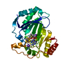
| ||||||||||||
|---|---|---|---|---|---|---|---|---|---|---|---|---|---|
| 1 |
| ||||||||||||
| Unit cell |
|
-Components
-Protein , 1 types, 1 molecules A
| #1: Protein | Mass: 28237.273 Da / Num. of mol.: 1 Mutation: S1E, E53K, A93E, R111H, Q139K, E151R, K154Q, N158E, K222E, K225R, K226A, E230R Source method: isolated from a genetically manipulated source Details: Computationally redesigned for reproducible crystallization Source: (gene. exp.) Details (production host): Kanamycin-resistant, cleavable N-terminal 10xHis-SUMO tag Production host: References: UniProt: P0A9N4, [formate-C-acetyltransferase]-activating enzyme |
|---|
-Non-polymers , 5 types, 258 molecules 


| #2: Chemical | ChemComp-SF4 /  Mass: 351.640 Da / Num. of mol.: 1 / Source method: obtained synthetically / Formula: Fe4S4 / Feature type: SUBJECT OF INVESTIGATION Mass: 351.640 Da / Num. of mol.: 1 / Source method: obtained synthetically / Formula: Fe4S4 / Feature type: SUBJECT OF INVESTIGATION | ||||
|---|---|---|---|---|---|
| #3: Chemical | ChemComp-SAM /  Mass: 398.437 Da / Num. of mol.: 1 / Source method: obtained synthetically / Formula: C15H22N6O5S / Feature type: SUBJECT OF INVESTIGATION Mass: 398.437 Da / Num. of mol.: 1 / Source method: obtained synthetically / Formula: C15H22N6O5S / Feature type: SUBJECT OF INVESTIGATION | ||||
| #4: Chemical |  Mass: 39.098 Da / Num. of mol.: 2 / Source method: obtained synthetically / Formula: K / Feature type: SUBJECT OF INVESTIGATION Mass: 39.098 Da / Num. of mol.: 2 / Source method: obtained synthetically / Formula: K / Feature type: SUBJECT OF INVESTIGATION#5: Chemical | ChemComp-CL / |  Mass: 35.453 Da / Num. of mol.: 1 / Source method: obtained synthetically / Formula: Cl Mass: 35.453 Da / Num. of mol.: 1 / Source method: obtained synthetically / Formula: Cl#6: Water | ChemComp-HOH / | Mass: 18.015 Da / Num. of mol.: 253 / Source method: isolated from a natural source / Formula: H2O |
-Details
| Has ligand of interest | Y |
|---|
-Experimental details
-Experiment
| Experiment | Method: X-RAY DIFFRACTION / Number of used crystals: 1 |
|---|
- Sample preparation
| Crystal | Density Matthews: 2.03 Å3/Da / Density % sol: 39.44 % / Description: rod-like |
|---|---|
| Crystal grow | Temperature: 300 K / Method: vapor diffusion, hanging drop / pH: 9 Details: 4 uL of protein (10 mg/mL PFL-AE-CCR8 in 12.5 mM HEPES, 200 mM KCl, 3.5 mM SAM, 5.0 mM WT 7-mer PFL peptide, and 2.5 mM DTT) with 1 uL of crystallization reservoir solution (28% PEG 3350, ...Details: 4 uL of protein (10 mg/mL PFL-AE-CCR8 in 12.5 mM HEPES, 200 mM KCl, 3.5 mM SAM, 5.0 mM WT 7-mer PFL peptide, and 2.5 mM DTT) with 1 uL of crystallization reservoir solution (28% PEG 3350, 100 mM glycine, pH 9.0) in hanging drop format over 50 uL of crystallization reservoir solution |
-Data collection
| Diffraction | Mean temperature: 100 K / Serial crystal experiment: N |
|---|---|
| Diffraction source | Source: SYNCHROTRON / Site: APS / Beamline: 24-ID-C / Wavelength: 0.9791 Å |
| Detector | Type: DECTRIS PILATUS 6M-F / Detector: PIXEL / Date: Feb 22, 2017 |
| Radiation | Protocol: SINGLE WAVELENGTH / Monochromatic (M) / Laue (L): M / Scattering type: x-ray |
| Radiation wavelength | Wavelength: 0.9791 Å / Relative weight: 1 |
| Reflection | Resolution: 1.46→41.77 Å / Num. obs: 39365 / % possible obs: 96.88 % / Redundancy: 7.6 % / Biso Wilson estimate: 16.31 Å2 / CC1/2: 0.999 / CC star: 1 / Rmerge(I) obs: 0.05246 / Rpim(I) all: 0.01912 / Rrim(I) all: 0.056 / Net I/σ(I): 19.76 |
| Reflection shell | Resolution: 1.46→1.512 Å / Redundancy: 7.7 % / Rmerge(I) obs: 0.2999 / Mean I/σ(I) obs: 3.66 / Num. unique obs: 3661 / CC1/2: 0.959 / CC star: 0.989 / Rpim(I) all: 0.1104 / Rrim(I) all: 0.3205 / % possible all: 91.96 |
- Processing
| Software |
| |||||||||||||||||||||||||||||||||||||||||||||||||||||||||||||||||||||||||||||||||||||||||||||||||||||||||||||||||||||||||||||
|---|---|---|---|---|---|---|---|---|---|---|---|---|---|---|---|---|---|---|---|---|---|---|---|---|---|---|---|---|---|---|---|---|---|---|---|---|---|---|---|---|---|---|---|---|---|---|---|---|---|---|---|---|---|---|---|---|---|---|---|---|---|---|---|---|---|---|---|---|---|---|---|---|---|---|---|---|---|---|---|---|---|---|---|---|---|---|---|---|---|---|---|---|---|---|---|---|---|---|---|---|---|---|---|---|---|---|---|---|---|---|---|---|---|---|---|---|---|---|---|---|---|---|---|---|---|---|
| Refinement | Method to determine structure: MOLECULAR REPLACEMENT / Resolution: 1.46→41.77 Å / SU ML: 0.1297 / Cross valid method: FREE R-VALUE / σ(F): 1.37 / Phase error: 14.752 Stereochemistry target values: GeoStd + Monomer Library + CDL v1.2
| |||||||||||||||||||||||||||||||||||||||||||||||||||||||||||||||||||||||||||||||||||||||||||||||||||||||||||||||||||||||||||||
| Solvent computation | Shrinkage radii: 0.9 Å / VDW probe radii: 1.1 Å / Solvent model: FLAT BULK SOLVENT MODEL | |||||||||||||||||||||||||||||||||||||||||||||||||||||||||||||||||||||||||||||||||||||||||||||||||||||||||||||||||||||||||||||
| Displacement parameters | Biso mean: 24.39 Å2 | |||||||||||||||||||||||||||||||||||||||||||||||||||||||||||||||||||||||||||||||||||||||||||||||||||||||||||||||||||||||||||||
| Refinement step | Cycle: LAST / Resolution: 1.46→41.77 Å
| |||||||||||||||||||||||||||||||||||||||||||||||||||||||||||||||||||||||||||||||||||||||||||||||||||||||||||||||||||||||||||||
| Refine LS restraints |
| |||||||||||||||||||||||||||||||||||||||||||||||||||||||||||||||||||||||||||||||||||||||||||||||||||||||||||||||||||||||||||||
| LS refinement shell |
| |||||||||||||||||||||||||||||||||||||||||||||||||||||||||||||||||||||||||||||||||||||||||||||||||||||||||||||||||||||||||||||
| Refinement TLS params. | Method: refined / Refine-ID: X-RAY DIFFRACTION
| |||||||||||||||||||||||||||||||||||||||||||||||||||||||||||||||||||||||||||||||||||||||||||||||||||||||||||||||||||||||||||||
| Refinement TLS group | Refine-ID: X-RAY DIFFRACTION / Auth asym-ID: A / Label asym-ID: A
|
