 Movie
Movie Controller

[English] 日本語
 Yorodumi
- PDB-8frc: Mouse acidic mammalian chitinase, catalytic domain in complex wit... -
+ Open data

- Basic information
| Entry | Database: PDB / ID: 8frc | |||||||||
|---|---|---|---|---|---|---|---|---|---|---|
| Title | Mouse acidic mammalian chitinase, catalytic domain in complex with N,N'-diacetylchitobiose at pH 4.91 | |||||||||
 Components Components | Acidic mammalian chitinase | |||||||||
 Keywords Keywords | HYDROLASE / GH18 chitinase / acidic mammalian chitinase | |||||||||
| Function / homology |  Function and homology information Function and homology informationDigestion of dietary carbohydrate / production of molecular mediator involved in inflammatory response / chitinase activity / endochitinase activity / chitinase / chitin catabolic process / chitin binding / polysaccharide catabolic process / immune system process / positive regulation of chemokine production ...Digestion of dietary carbohydrate / production of molecular mediator involved in inflammatory response / chitinase activity / endochitinase activity / chitinase / chitin catabolic process / chitin binding / polysaccharide catabolic process / immune system process / positive regulation of chemokine production / apoptotic process / extracellular space / cytoplasm Similarity search - Function | |||||||||
| Biological species |  | |||||||||
| Method |  X-RAY DIFFRACTION / SYNCHROTRON / MOLECULAR REPLACEMENT / Resolution: 1.92 Å X-RAY DIFFRACTION / SYNCHROTRON / MOLECULAR REPLACEMENT / Resolution: 1.92 Å | |||||||||
 Authors Authors | Diaz, R.E. / Fraser, J.S. | |||||||||
| Funding support |  United States, 2items United States, 2items
| |||||||||
 Citation Citation | Journal: Biorxiv / Year: 2024 Title: Structural characterization of ligand binding and pH-specific enzymatic activity of mouse Acidic Mammalian Chitinase. Authors: Diaz, R.E. / Ecker, A.K. / Correy, G.J. / Asthana, P. / Young, I.D. / Faust, B. / Thompson, M.C. / Seiple, I.B. / Van Dyken, S.J. / Locksley, R.M. / Fraser, J.S. | |||||||||
| History |
|
- Structure visualization
| Structure viewer | Molecule: MolmilJmol/JSmol |
|---|
- Downloads & links
-Download
| PDBx/mmCIF format | 8frc.cif.gz | 376.1 KB | Display | PDBx/mmCIF format |
|---|---|---|---|---|
| PDB format | pdb8frc.ent.gz | 250.6 KB | Display | PDB format |
| PDBx/mmJSON format | 8frc.json.gz | Tree view | PDBx/mmJSON format | |
| Others |  Other downloads Other downloads |
-Validation report
| Summary document | 8frc_validation.pdf.gz | 1.7 MB | Display | wwPDB validaton report |
|---|---|---|---|---|
| Full document | 8frc_full_validation.pdf.gz | 1.7 MB | Display | |
| Data in XML | 8frc_validation.xml.gz | 35.6 KB | Display | |
| Data in CIF | 8frc_validation.cif.gz | 55 KB | Display | |
| Arichive directory | https://data.pdbj.org/pub/pdb/validation_reports/fr/8frcftp://data.pdbj.org/pub/pdb/validation_reports/fr/8frc | HTTPS FTP |
-Related structure data
| Related structure data | 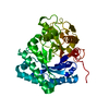 8fg5C 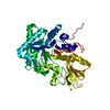 8fg7C 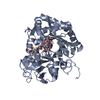 8fr9C 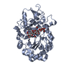 8fraC 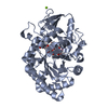 8frbC 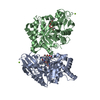 8frdC 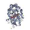 8frgC 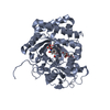 8gcaC C: citing same article ( |
|---|---|
| Similar structure data |
-Links
 PDBj
PDBj- Assembly
| Deposited unit | 
| ||||||||||||
|---|---|---|---|---|---|---|---|---|---|---|---|---|---|
| 1 | 
| ||||||||||||
| 2 | 
| ||||||||||||
| Unit cell |
|
-Components
| #1: Protein | Mass: 44621.992 Da / Num. of mol.: 2 Source method: isolated from a genetically manipulated source Source: (gene. exp.)  Cricetulus griseus (Chinese hamster) / References: UniProt: Q91XA9, chitinase Cricetulus griseus (Chinese hamster) / References: UniProt: Q91XA9, chitinase#2: Polysaccharide | 2-acetamido-2-deoxy-beta-D-glucopyranose-(1-4)-2-acetamido-2-deoxy-beta-D-glucopyranose #3: Water | ChemComp-HOH / |  Mass: 18.015 Da / Num. of mol.: 740 / Source method: isolated from a natural source / Formula: H2O Mass: 18.015 Da / Num. of mol.: 740 / Source method: isolated from a natural source / Formula: H2OHas ligand of interest | Y | Has protein modification | Y | |
|---|
-Experimental details
-Experiment
| Experiment | Method: X-RAY DIFFRACTION / Number of used crystals: 1 |
|---|
- Sample preparation
| Crystal | Density Matthews: 1.94 Å3/Da / Density % sol: 36.47 % |
|---|---|
| Crystal grow | Temperature: 293.15 K / Method: vapor diffusion, hanging drop / pH: 4.91 Details: 11 mg/mL AMCase catalytic domain; 20% w/v PEG 6000; 0.1 M Sodium Acetate pH 4.91; 0.2 M Magnesium Chloride; 29.00 mM GlcNAc2 |
-Data collection
| Diffraction | Mean temperature: 100 K / Serial crystal experiment: N |
|---|---|
| Diffraction source | Source: SYNCHROTRON / Site: ALS / Beamline: 8.3.1 / Wavelength: 1.11578 Å |
| Detector | Type: DECTRIS PILATUS3 6M / Detector: PIXEL / Date: Jun 15, 2021 |
| Radiation | Protocol: SINGLE WAVELENGTH / Monochromatic (M) / Laue (L): M / Scattering type: x-ray |
| Radiation wavelength | Wavelength: 1.11578 Å / Relative weight: 1 |
| Reflection | Resolution: 1.92→105.12 Å / Num. obs: 53659 / % possible obs: 100 % / Redundancy: 6.3 % / Biso Wilson estimate: 13.44 Å2 / CC1/2: 0.994 / Rmerge(I) obs: 0.153 / Rpim(I) all: 0.066 / Rrim(I) all: 0.167 / Net I/σ(I): 7.7 |
| Reflection shell | Resolution: 1.92→1.95 Å / Redundancy: 6.5 % / Rmerge(I) obs: 0.568 / Mean I/σ(I) obs: 3 / Num. unique obs: 2576 / CC1/2: 0.868 / Rpim(I) all: 0.239 / Rrim(I) all: 0.617 / % possible all: 98.6 |
- Processing
| Software |
| |||||||||||||||||||||||||||||||||||||||||||||||||||||||||||||||||||||||||||||||||||||||||||||||||||||||||||||||||||||||||||||||||||||||||||||||||||
|---|---|---|---|---|---|---|---|---|---|---|---|---|---|---|---|---|---|---|---|---|---|---|---|---|---|---|---|---|---|---|---|---|---|---|---|---|---|---|---|---|---|---|---|---|---|---|---|---|---|---|---|---|---|---|---|---|---|---|---|---|---|---|---|---|---|---|---|---|---|---|---|---|---|---|---|---|---|---|---|---|---|---|---|---|---|---|---|---|---|---|---|---|---|---|---|---|---|---|---|---|---|---|---|---|---|---|---|---|---|---|---|---|---|---|---|---|---|---|---|---|---|---|---|---|---|---|---|---|---|---|---|---|---|---|---|---|---|---|---|---|---|---|---|---|---|---|---|---|
| Refinement | Method to determine structure: MOLECULAR REPLACEMENT / Resolution: 1.92→70.93 Å / SU ML: 0.1678 / Cross valid method: FREE R-VALUE / σ(F): 2 / Phase error: 16.6686 Stereochemistry target values: GeoStd + Monomer Library + CDL v1.2
| |||||||||||||||||||||||||||||||||||||||||||||||||||||||||||||||||||||||||||||||||||||||||||||||||||||||||||||||||||||||||||||||||||||||||||||||||||
| Solvent computation | Shrinkage radii: 0.9 Å / VDW probe radii: 1.1 Å / Solvent model: FLAT BULK SOLVENT MODEL | |||||||||||||||||||||||||||||||||||||||||||||||||||||||||||||||||||||||||||||||||||||||||||||||||||||||||||||||||||||||||||||||||||||||||||||||||||
| Displacement parameters | Biso mean: 14.55 Å2 | |||||||||||||||||||||||||||||||||||||||||||||||||||||||||||||||||||||||||||||||||||||||||||||||||||||||||||||||||||||||||||||||||||||||||||||||||||
| Refinement step | Cycle: LAST / Resolution: 1.92→70.93 Å
| |||||||||||||||||||||||||||||||||||||||||||||||||||||||||||||||||||||||||||||||||||||||||||||||||||||||||||||||||||||||||||||||||||||||||||||||||||
| Refine LS restraints |
| |||||||||||||||||||||||||||||||||||||||||||||||||||||||||||||||||||||||||||||||||||||||||||||||||||||||||||||||||||||||||||||||||||||||||||||||||||
| LS refinement shell |
|
