 Movie
Movie Controller


+ Open data

- Basic information
| Entry | Database: PDB / ID: 8fov | ||||||
|---|---|---|---|---|---|---|---|
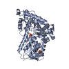
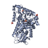
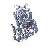
| Title | AbeH (Tryptophan-5-halogenase) bound to FAD and Cl | ||||||
 Components Components | Tryptophan 5-halogenase | ||||||
 Keywords Keywords | OXIDOREDUCTASE / halogenase / flavin | ||||||
| Function / homology |  Function and homology information Function and homology information | ||||||
| Biological species |  uncultured bacterium (environmental samples) uncultured bacterium (environmental samples) | ||||||
| Method |  X-RAY DIFFRACTION / SYNCHROTRON / MOLECULAR REPLACEMENT / molecular replacement / Resolution: 1.86 Å X-RAY DIFFRACTION / SYNCHROTRON / MOLECULAR REPLACEMENT / molecular replacement / Resolution: 1.86 Å | ||||||
 Authors Authors | Ashaduzzaman, M. / Bellizzi, J.J. | ||||||
| Funding support |  United States, 1items United States, 1items
| ||||||
 Citation Citation | Journal: Acs Omega / Year: 2025 Title: Crystallographic and Thermodynamic Evidence of Negative Coupling in the Flavin-Dependent Tryptophan Halogenases AbeH and BorH. Authors: Ashaduzzaman, M. / Lingkon, K. / De Silva, A.J. / Bellizzi 3rd, J.J. #1: Journal: Biorxiv / Year: 2023 Title: Crystallographic and thermodynamic evidence of negative cooperativity of flavin and tryptophan binding in the flavin-dependent halogenases AbeH and BorH. Authors: Ashaduzzaman, M. / Lingkon, K. / De Silva, A.J. / Bellizzi, J.J. | ||||||
| History |
|
- Structure visualization
| Structure viewer | Molecule: MolmilJmol/JSmol |
|---|
- Downloads & links
-Download
| PDBx/mmCIF format | 8fov.cif.gz | 447.8 KB | Display | PDBx/mmCIF format |
|---|---|---|---|---|
| PDB format | pdb8fov.ent.gz | 349.1 KB | Display | PDB format |
| PDBx/mmJSON format | 8fov.json.gz | Tree view | PDBx/mmJSON format | |
| Others |  Other downloads Other downloads |
-Validation report
| Summary document | 8fov_validation.pdf.gz | 939.9 KB | Display | wwPDB validaton report |
|---|---|---|---|---|
| Full document | 8fov_full_validation.pdf.gz | 945.3 KB | Display | |
| Data in XML | 8fov_validation.xml.gz | 44.3 KB | Display | |
| Data in CIF | 8fov_validation.cif.gz | 65.8 KB | Display | |
| Arichive directory | https://data.pdbj.org/pub/pdb/validation_reports/fo/8fovftp://data.pdbj.org/pub/pdb/validation_reports/fo/8fov | HTTPS FTP |
-Related structure data

-Links
 PDBj
PDBj- Assembly
| Deposited unit | 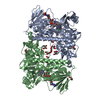
| ||||||||||
|---|---|---|---|---|---|---|---|---|---|---|---|
| 1 | 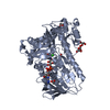
| ||||||||||
| 2 | 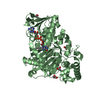
| ||||||||||
| Unit cell |
|
-Components
-Protein , 1 types, 2 molecules AB
| #1: Protein | Mass: 58350.500 Da / Num. of mol.: 2 Source method: isolated from a genetically manipulated source Details: Contains extra Proline at N-terminus (residue 0) remaining from cleavage of linker with N-terminal His-tag. Source: (gene. exp.) uncultured bacterium (environmental samples)Gene: abeH / Production host: |
|---|
-Non-polymers , 5 types, 825 molecules 


| #2: Chemical |  Mass: 785.550 Da / Num. of mol.: 2 / Source method: obtained synthetically / Formula: C27H33N9O15P2 / Feature type: SUBJECT OF INVESTIGATION / Comment: FAD*YM Mass: 785.550 Da / Num. of mol.: 2 / Source method: obtained synthetically / Formula: C27H33N9O15P2 / Feature type: SUBJECT OF INVESTIGATION / Comment: FAD*YM#3: Chemical |  Mass: 35.453 Da / Num. of mol.: 2 / Source method: obtained synthetically / Formula: Cl Mass: 35.453 Da / Num. of mol.: 2 / Source method: obtained synthetically / Formula: Cl#4: Chemical | ChemComp-GOL /  Mass: 92.094 Da / Num. of mol.: 8 / Source method: obtained synthetically / Formula: C3H8O3 Mass: 92.094 Da / Num. of mol.: 8 / Source method: obtained synthetically / Formula: C3H8O3#5: Chemical | ChemComp-ACT /  Mass: 59.044 Da / Num. of mol.: 4 / Source method: obtained synthetically / Formula: C2H3O2 Mass: 59.044 Da / Num. of mol.: 4 / Source method: obtained synthetically / Formula: C2H3O2#6: Water | ChemComp-HOH / | Mass: 18.015 Da / Num. of mol.: 809 / Source method: isolated from a natural source / Formula: H2O |
|---|
-Details
| Has ligand of interest | Y |
|---|---|
| Has protein modification | N |
-Experimental details
-Experiment
| Experiment | Method: X-RAY DIFFRACTION / Number of used crystals: 1 |
|---|
- Sample preparation
| Crystal | Density Matthews: 2.37 Å3/Da / Density % sol: 48.16 % |
|---|---|
| Crystal grow | Temperature: 293 K / Method: vapor diffusion, hanging drop / pH: 6.2 Details: Drop:1:1 ratio of reservoir solution and (50uM AbeH/25 mM FAD in 20 mM HEPES pH 7.2 and 35 mM sodium citrate); Reservoir (500 uL): 0.1 M Bis-Tris pH 6.2, 0.2 M magnesium acetate, 11% (v/v) PEG 10,000 |
-Data collection
| Diffraction | Mean temperature: 100 K / Serial crystal experiment: N |
|---|---|
| Diffraction source | Source: SYNCHROTRON / Site: APS / Beamline: 21-ID-F / Wavelength: 0.97872 Å |
| Detector | Type: RAYONIX MX-300 / Detector: CCD / Date: Nov 7, 2018 |
| Radiation | Protocol: SINGLE WAVELENGTH / Monochromatic (M) / Laue (L): M / Scattering type: x-ray |
| Radiation wavelength | Wavelength: 0.97872 Å / Relative weight: 1 |
| Reflection | Resolution: 1.86→56.14 Å / Num. obs: 93637 / % possible obs: 99.63 % / Observed criterion σ(I): 2 / Redundancy: 2 % / Biso Wilson estimate: 23.36 Å2 / CC1/2: 0.99 / CC star: 0.998 / Rmerge(I) obs: 0.06443 / Rpim(I) all: 0.06443 / Rrim(I) all: 0.09112 / Net I/σ(I): 7.1 |
| Reflection shell | Resolution: 1.86→1.926 Å / Redundancy: 2 % / Rmerge(I) obs: 0.3797 / Mean I/σ(I) obs: 2.06 / Num. unique obs: 9276 / CC1/2: 0.708 / CC star: 0.911 / Rpim(I) all: 0.3797 / Rrim(I) all: 0.537 / % possible all: 99.96 |
-Phasing
| Phasing | Method: molecular replacement |
|---|
- Processing
| Software |
| |||||||||||||||||||||||||||||||||||||||||||||||||
|---|---|---|---|---|---|---|---|---|---|---|---|---|---|---|---|---|---|---|---|---|---|---|---|---|---|---|---|---|---|---|---|---|---|---|---|---|---|---|---|---|---|---|---|---|---|---|---|---|---|---|
| Refinement | Method to determine structure: MOLECULAR REPLACEMENT / Resolution: 1.86→56.14 Å / SU ML: 0.1911 / Cross valid method: FREE R-VALUE / σ(F): 0.6 / Phase error: 20.1232 Stereochemistry target values: GeoStd + Monomer Library + CDL v1.2
| |||||||||||||||||||||||||||||||||||||||||||||||||
| Solvent computation | Shrinkage radii: 0.9 Å / VDW probe radii: 1.11 Å / Solvent model: FLAT BULK SOLVENT MODEL | |||||||||||||||||||||||||||||||||||||||||||||||||
| Displacement parameters | Biso mean: 29.53 Å2 | |||||||||||||||||||||||||||||||||||||||||||||||||
| Refinement step | Cycle: LAST / Resolution: 1.86→56.14 Å
| |||||||||||||||||||||||||||||||||||||||||||||||||
| Refine LS restraints |
| |||||||||||||||||||||||||||||||||||||||||||||||||
| LS refinement shell |
|
