Deposited unit
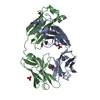
A: H9 immunoglobulin light chain
B: H9 immunoglobulin light chain
C: H9 immunoglobulin light chain
D: H9 immunoglobulin light chain
G: H9 immunoglobulin light chain
H: H9 immunoglobulin light chain
I: H9 immunoglobulin light chain
J: H9 immunoglobulin light chain
E: H9 immunoglobulin light chain
F: H9 immunoglobulin light chain
K: H9 immunoglobulin light chain
L: H9 immunoglobulin light chain
hetero molecules Summary Component details
Theoretical mass Number of molelcules Total (without water) 273,268 24 Polymers 270,596 12 Non-polymers 2,672 12 Water 25,041 1390
1
A: H9 immunoglobulin light chain
B: H9 immunoglobulin light chain
hetero molecules Summary Component details Symmetry operations Calculated values
Theoretical mass Number of molelcules Total (without water) 45,640 5 Polymers 45,099 2 Non-polymers 540 3 Water 36 2
Type Name Symmetry operation Number identity operation 1_555 x,y,z 1
Buried area 3630 Å2 ΔGint -24 kcal/mol Surface area 19620 Å2 Method
2
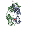
C: H9 immunoglobulin light chain
D: H9 immunoglobulin light chain
hetero molecules Summary Component details Symmetry operations Calculated values
Theoretical mass Number of molelcules Total (without water) 45,450 3 Polymers 45,099 2 Non-polymers 350 1 Water 36 2
Type Name Symmetry operation Number identity operation 1_555 x,y,z 1
Buried area 3580 Å2 ΔGint -25 kcal/mol Surface area 19730 Å2 Method
3
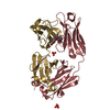
G: H9 immunoglobulin light chain
H: H9 immunoglobulin light chain
hetero molecules Summary Component details Symmetry operations Calculated values
Theoretical mass Number of molelcules Total (without water) 45,640 5 Polymers 45,099 2 Non-polymers 540 3 Water 36 2
Type Name Symmetry operation Number identity operation 1_555 x,y,z 1
Buried area 3420 Å2 ΔGint -21 kcal/mol Surface area 19160 Å2 Method
4
I: H9 immunoglobulin light chain
J: H9 immunoglobulin light chain
hetero molecules Summary Component details Symmetry operations Calculated values
Theoretical mass Number of molelcules Total (without water) 45,450 3 Polymers 45,099 2 Non-polymers 350 1 Water 36 2
Type Name Symmetry operation Number identity operation 1_555 x,y,z 1
Buried area 3500 Å2 ΔGint -21 kcal/mol Surface area 19510 Å2 Method
5
E: H9 immunoglobulin light chain
F: H9 immunoglobulin light chain
hetero molecules Summary Component details Symmetry operations Calculated values
Theoretical mass Number of molelcules Total (without water) 45,545 4 Polymers 45,099 2 Non-polymers 445 2 Water 36 2
Type Name Symmetry operation Number identity operation 1_555 x,y,z 1
Buried area 3500 Å2 ΔGint -21 kcal/mol Surface area 19220 Å2 Method
6
K: H9 immunoglobulin light chain
L: H9 immunoglobulin light chain
hetero molecules Summary Component details Symmetry operations Calculated values
Theoretical mass Number of molelcules Total (without water) 45,545 4 Polymers 45,099 2 Non-polymers 445 2 Water 36 2
Type Name Symmetry operation Number identity operation 1_555 x,y,z 1
Buried area 3470 Å2 ΔGint -22 kcal/mol Surface area 19590 Å2 Method
Unit cell Length a, b, c (Å) 63.141, 95.655, 125.918 Angle α, β, γ (deg.) 106.31, 92.93, 90.16 Int Tables number 1 Space group name H-M P1
 Movie
Movie Controller

 Yorodumi
 Open data
 Basic information
 Components
 Keywords
 Function and homology information
 Homo sapiens (human)
 X-RAY DIFFRACTION /
  Authors
 United States, 2items
  Citation
 Structure visualization
 Downloads & links
 Other downloads
 PDBj
PDBj

 Assembly


 Mass: 350.393 Da / Num. of mol.: 6 / Source method: obtained synthetically / Formula: C15H18N4O4S / Feature type: SUBJECT OF INVESTIGATION
 Mass: 94.971 Da / Num. of mol.: 6 / Source method: obtained synthetically / Formula: PO4
 Mass: 18.015 Da / Num. of mol.: 1390 / Source method: isolated from a natural source / Formula: H2O
 Sample preparation
 Processing