 Movie
Movie Controller

[English] 日本語
 Yorodumi
- PDB-8fnw: Structure of RdrA-RdrB complex from Escherichia coli RADAR defens... -
+ Open data

- Basic information
| Entry | Database: PDB / ID: 8fnw | |||||||||||||||
|---|---|---|---|---|---|---|---|---|---|---|---|---|---|---|---|---|
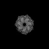
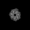
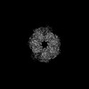
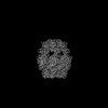
| Title | Structure of RdrA-RdrB complex from Escherichia coli RADAR defense system | |||||||||||||||
 Components Components |
| |||||||||||||||
 Keywords Keywords | IMMUNE SYSTEM / anti-phage defense / adenosine deaminase | |||||||||||||||
| Function / homology | deaminase activity / Adenosine/adenine deaminase / Metal-dependent hydrolase / P-loop containing nucleoside triphosphate hydrolase / Adenosine deaminase / Archaeal ATPase Function and homology information Function and homology information | |||||||||||||||
| Biological species |  | |||||||||||||||
| Method | ELECTRON MICROSCOPY / single particle reconstruction / cryo EM / Resolution: 6.73 Å | |||||||||||||||
 Authors Authors | Duncan-Lowey, B. / Johnson, A.G. / Rawson, S. / Mayer, M.L. / Kranzusch, P.J. | |||||||||||||||
| Funding support |  United States, 4items United States, 4items
| |||||||||||||||
 Citation Citation | Journal: Cell / Year: 2023 Title: Cryo-EM structure of the RADAR supramolecular anti-phage defense complex. Authors: Brianna Duncan-Lowey / Nitzan Tal / Alex G Johnson / Shaun Rawson / Megan L Mayer / Shany Doron / Adi Millman / Sarah Melamed / Taya Fedorenko / Assaf Kacen / Alexander Brandis / Tevie ...Authors: Brianna Duncan-Lowey / Nitzan Tal / Alex G Johnson / Shaun Rawson / Megan L Mayer / Shany Doron / Adi Millman / Sarah Melamed / Taya Fedorenko / Assaf Kacen / Alexander Brandis / Tevie Mehlman / Gil Amitai / Rotem Sorek / Philip J Kranzusch /  Abstract: RADAR is a two-protein bacterial defense system that was reported to defend against phage by "editing" messenger RNA. Here, we determine cryo-EM structures of the RADAR defense complex, revealing ...RADAR is a two-protein bacterial defense system that was reported to defend against phage by "editing" messenger RNA. Here, we determine cryo-EM structures of the RADAR defense complex, revealing RdrA as a heptameric, two-layered AAA+ ATPase and RdrB as a dodecameric, hollow complex with twelve surface-exposed deaminase active sites. RdrA and RdrB join to form a giant assembly up to 10 MDa, with RdrA docked as a funnel over the RdrB active site. Surprisingly, our structures reveal an RdrB active site that targets mononucleotides. We show that RdrB catalyzes ATP-to-ITP conversion in vitro and induces the massive accumulation of inosine mononucleotides during phage infection in vivo, limiting phage replication. Our results define ATP mononucleotide deamination as a determinant of RADAR immunity and reveal supramolecular assembly of a nucleotide-modifying machine as a mechanism of anti-phage defense. | |||||||||||||||
| History |
|
- Structure visualization
| Structure viewer | Molecule: MolmilJmol/JSmol |
|---|
- Downloads & links
-Download
| PDBx/mmCIF format | 8fnw.cif.gz | 2.5 MB | Display | PDBx/mmCIF format |
|---|---|---|---|---|
| PDB format | pdb8fnw.ent.gz | Display | PDB format | |
| PDBx/mmJSON format | 8fnw.json.gz | Tree view | PDBx/mmJSON format | |
| Others |  Other downloads Other downloads |
-Validation report
| Summary document | 8fnw_validation.pdf.gz | 1.8 MB | Display | wwPDB validaton report |
|---|---|---|---|---|
| Full document | 8fnw_full_validation.pdf.gz | 2.8 MB | Display | |
| Data in XML | 8fnw_validation.xml.gz | 524.9 KB | Display | |
| Data in CIF | 8fnw_validation.cif.gz | 750.8 KB | Display | |
| Arichive directory | https://data.pdbj.org/pub/pdb/validation_reports/fn/8fnwftp://data.pdbj.org/pub/pdb/validation_reports/fn/8fnw | HTTPS FTP |
-Related structure data
| Related structure data | 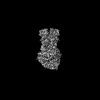 29328MC 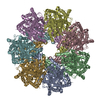 8fntC 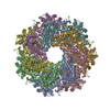 8fnuC 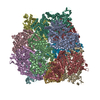 8fnvC M: map data used to model this data C: citing same article ( |
|---|---|
| Similar structure data |
-Links
 PDBj
PDBj

- Assembly
| Deposited unit | 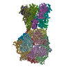
|
|---|---|
| 1 |
|
-Components
| #1: Protein | Mass: 92216.953 Da / Num. of mol.: 12 Source method: isolated from a genetically manipulated source Source: (gene. exp.) #2: Protein | Mass: 107226.594 Da / Num. of mol.: 7 Source method: isolated from a genetically manipulated source Source: (gene. exp.) #3: Chemical | ChemComp-ZN /   Mass: 65.409 Da / Num. of mol.: 12 / Source method: obtained synthetically / Formula: Zn Mass: 65.409 Da / Num. of mol.: 12 / Source method: obtained synthetically / Formula: ZnHas ligand of interest | N | |
|---|
-Experimental details
-Experiment
| Experiment | Method: ELECTRON MICROSCOPY |
|---|---|
| EM experiment | Aggregation state: PARTICLE / 3D reconstruction method: single particle reconstruction |
- Sample preparation
| Component | Name: Escherichia coli RdrA-RrdB complex / Type: COMPLEX / Entity ID: #1-#2 / Source: RECOMBINANT | ||||||||||||||||||||
|---|---|---|---|---|---|---|---|---|---|---|---|---|---|---|---|---|---|---|---|---|---|
| Molecular weight | Experimental value: NO | ||||||||||||||||||||
| Source (natural) | Organism: | ||||||||||||||||||||
| Source (recombinant) | Organism: | ||||||||||||||||||||
| Buffer solution | pH: 7.5 | ||||||||||||||||||||
| Buffer component |
| ||||||||||||||||||||
| Specimen | Embedding applied: NO / Shadowing applied: NO / Staining applied: NO / Vitrification applied: YES | ||||||||||||||||||||
| Vitrification | Cryogen name: ETHANE / Humidity: 100 % |
- Electron microscopy imaging
| Experimental equipment |  Model: Talos Arctica / Image courtesy: FEI Company |
|---|---|
| Microscopy | Model: FEI TALOS ARCTICA |
| Electron gun | Electron source:  FIELD EMISSION GUN / Accelerating voltage: 200 kV / Illumination mode: OTHER FIELD EMISSION GUN / Accelerating voltage: 200 kV / Illumination mode: OTHER |
| Electron lens | Mode: OTHER / Nominal defocus max: 3000 nm / Nominal defocus min: 500 nm |
| Image recording | Electron dose: 49 e/Å2 / Film or detector model: GATAN K3 (6k x 4k) |
- Processing
| Software |
| ||||||||||||||||||||||||
|---|---|---|---|---|---|---|---|---|---|---|---|---|---|---|---|---|---|---|---|---|---|---|---|---|---|
| CTF correction | Type: NONE | ||||||||||||||||||||||||
| 3D reconstruction | Resolution: 6.73 Å / Resolution method: FSC 0.143 CUT-OFF / Num. of particles: 9236 / Symmetry type: POINT | ||||||||||||||||||||||||
| Refinement | Cross valid method: NONE Stereochemistry target values: GeoStd + Monomer Library + CDL v1.2 | ||||||||||||||||||||||||
| Displacement parameters | Biso mean: 269.44 Å2 | ||||||||||||||||||||||||
| Refine LS restraints |
|
