 Movie
Movie Controller

[English] 日本語
 Yorodumi
- PDB-8fn5: Structure of the truncated catalytic domain of Streptococcus muta... -
+ Open data

- Basic information
| Entry | Database: PDB / ID: 8fn5 | ||||||
|---|---|---|---|---|---|---|---|
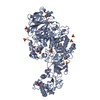
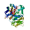
| Title | Structure of the truncated catalytic domain of Streptococcus mutans GtfD | ||||||
 Components Components | Glucosyltransferase-S | ||||||
 Keywords Keywords | TRANSFERASE / GtfD / GTF-S / soluble 1 / 6-linked alpha glucans / biofilm / glucansucrase | ||||||
| Function / homology |  Function and homology information Function and homology informationdextransucrase activity / dextransucrase / glucan biosynthetic process / glucosyltransferase activity / extracellular region Similarity search - Function | ||||||
| Biological species |  Streptococcus mutans (bacteria) Streptococcus mutans (bacteria) | ||||||
| Method |  X-RAY DIFFRACTION / SYNCHROTRON / MOLECULAR REPLACEMENT / Resolution: 1.92 Å X-RAY DIFFRACTION / SYNCHROTRON / MOLECULAR REPLACEMENT / Resolution: 1.92 Å | ||||||
 Authors Authors | Schormann, N. / Deivanayagam, C. | ||||||
| Funding support | 1items
| ||||||
 Citation Citation | Journal: Acta Crystallogr.,Sect.F / Year: 2023 Title: The catalytic domains of Streptococcus mutans glucosyltransferases: a structural analysis. Authors: Schormann, N. / Patel, M. / Thannickal, L. / Purushotham, S. / Wu, R. / Mieher, J.L. / Wu, H. / Deivanayagam, C. | ||||||
| History |
|
- Structure visualization
| Structure viewer | Molecule: MolmilJmol/JSmol |
|---|
- Downloads & links
-Download
| PDBx/mmCIF format | 8fn5.cif.gz | 248.5 KB | Display | PDBx/mmCIF format |
|---|---|---|---|---|
| PDB format | pdb8fn5.ent.gz | 196.2 KB | Display | PDB format |
| PDBx/mmJSON format | 8fn5.json.gz | Tree view | PDBx/mmJSON format | |
| Others |  Other downloads Other downloads |
-Validation report
| Arichive directory | https://data.pdbj.org/pub/pdb/validation_reports/fn/8fn5ftp://data.pdbj.org/pub/pdb/validation_reports/fn/8fn5 | HTTPS FTP |
|---|
-Related structure data


-Links
 PDBj
PDBj
- Assembly
| Deposited unit | 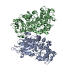
| ||||||||
|---|---|---|---|---|---|---|---|---|---|
| 1 | 
| ||||||||
| 2 | 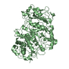
| ||||||||
| Unit cell |
|
-Components
| #1: Protein | Mass: 70604.227 Da / Num. of mol.: 2 Source method: isolated from a genetically manipulated source Details: Crystal structure shows a truncated catalytic domain of GtfD. The initial purified construct stretches from 248 to 1090. Proteolysis occurred during crystallization, and resulted in the ...Details: Crystal structure shows a truncated catalytic domain of GtfD. The initial purified construct stretches from 248 to 1090. Proteolysis occurred during crystallization, and resulted in the residue range of 423-1054. Source: (gene. exp.) Streptococcus mutans (bacteria) / Gene: gtfD, SMU_910 / Plasmid: pET-21a / Production host: #2: Water | ChemComp-HOH / |  Mass: 18.015 Da / Num. of mol.: 391 / Source method: isolated from a natural source / Formula: H2O Mass: 18.015 Da / Num. of mol.: 391 / Source method: isolated from a natural source / Formula: H2O |
|---|
-Experimental details
-Experiment
| Experiment | Method: X-RAY DIFFRACTION / Number of used crystals: 1 |
|---|
- Sample preparation
| Crystal | Density Matthews: 2.36 Å3/Da / Density % sol: 47.85 % |
|---|---|
| Crystal grow | Temperature: 295 K / Method: vapor diffusion, hanging drop / pH: 5.8 / Details: 26% PEG 2000MME, 0.1 M Bis-Tris |
-Data collection
| Diffraction | Mean temperature: 100 K / Serial crystal experiment: N |
|---|---|
| Diffraction source | Source: SYNCHROTRON / Site: APS  / Beamline: 22-ID / Wavelength: 1 Å / Beamline: 22-ID / Wavelength: 1 Å |
| Detector | Type: DECTRIS EIGER X 16M / Detector: PIXEL / Date: Jun 13, 2021 |
| Radiation | Protocol: SINGLE WAVELENGTH / Monochromatic (M) / Laue (L): M / Scattering type: x-ray |
| Radiation wavelength | Wavelength: 1 Å / Relative weight: 1 |
| Reflection | Resolution: 1.92→86.83 Å / Num. obs: 97783 / % possible obs: 95.6 % / Redundancy: 3.6 % / CC1/2: 0.996 / Rmerge(I) obs: 0.1 / Rpim(I) all: 0.057 / Rrim(I) all: 0.116 / Χ2: 1.01 / Net I/σ(I): 7.7 |
| Reflection shell | Resolution: 1.92→1.95 Å / Redundancy: 3.7 % / Rmerge(I) obs: 0.797 / Mean I/σ(I) obs: 1.7 / Num. unique obs: 4973 / CC1/2: 0.657 / CC star: 0.891 / Rpim(I) all: 0.469 / Rrim(I) all: 0.93 / Χ2: 1.05 / % possible all: 98.5 |
- Processing
| Software |
| ||||||||||||||||||||||||||||||||||||||||||||||||||||||||||||
|---|---|---|---|---|---|---|---|---|---|---|---|---|---|---|---|---|---|---|---|---|---|---|---|---|---|---|---|---|---|---|---|---|---|---|---|---|---|---|---|---|---|---|---|---|---|---|---|---|---|---|---|---|---|---|---|---|---|---|---|---|---|
| Refinement | Method to determine structure: MOLECULAR REPLACEMENT / Resolution: 1.92→86.83 Å / Cor.coef. Fo:Fc: 0.945 / Cor.coef. Fo:Fc free: 0.935 / SU B: 5.85 / SU ML: 0.151 / Cross valid method: THROUGHOUT / σ(F): 0 / ESU R: 0.191 / ESU R Free: 0.161 / Stereochemistry target values: MAXIMUM LIKELIHOOD Details: HYDROGENS HAVE BEEN ADDED IN THE RIDING POSITIONS U VALUES : REFINED INDIVIDUALLY
| ||||||||||||||||||||||||||||||||||||||||||||||||||||||||||||
| Solvent computation | Ion probe radii: 0.8 Å / Shrinkage radii: 0.8 Å / VDW probe radii: 1.2 Å / Solvent model: MASK | ||||||||||||||||||||||||||||||||||||||||||||||||||||||||||||
| Displacement parameters | Biso max: 99.67 Å2 / Biso mean: 31.257 Å2 / Biso min: 15.87 Å2
| ||||||||||||||||||||||||||||||||||||||||||||||||||||||||||||
| Refinement step | Cycle: final / Resolution: 1.92→86.83 Å
| ||||||||||||||||||||||||||||||||||||||||||||||||||||||||||||
| Refine LS restraints |
| ||||||||||||||||||||||||||||||||||||||||||||||||||||||||||||
| LS refinement shell | Resolution: 1.92→1.97 Å / Rfactor Rfree error: 0 / Total num. of bins used: 20
|
