 Movie
Movie Controller

[English] 日本語
 Yorodumi
- PDB-8fj9: Structure of the catalytic domain of Streptococcus mutans GtfB in... -
+ Open data

- Basic information
| Entry | Database: PDB / ID: 8fj9 | ||||||
|---|---|---|---|---|---|---|---|
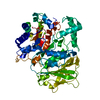
| Title | Structure of the catalytic domain of Streptococcus mutans GtfB in tetragonal space group P4322 | ||||||
 Components Components | Glucosyltransferase-I | ||||||
 Keywords Keywords | TRANSFERASE / GtfB / GTF-I / insoluble 1 / 3-linked alpha Glucans / Biofilm / Glucansucrase | ||||||
| Function / homology |  Function and homology information Function and homology informationdextransucrase activity / dextransucrase / glucan biosynthetic process / glucosyltransferase activity / extracellular region Similarity search - Function | ||||||
| Biological species |  Streptococcus mutans (bacteria) Streptococcus mutans (bacteria) | ||||||
| Method |  X-RAY DIFFRACTION / SYNCHROTRON / MOLECULAR REPLACEMENT / Resolution: 2.5 Å X-RAY DIFFRACTION / SYNCHROTRON / MOLECULAR REPLACEMENT / Resolution: 2.5 Å | ||||||
 Authors Authors | Schormann, N. / Deivanayagam, C. | ||||||
| Funding support | 1items
| ||||||
 Citation Citation | Journal: Acta Crystallogr.,Sect.F / Year: 2023 Title: The catalytic domains of Streptococcus mutans glucosyltransferases: a structural analysis. Authors: Schormann, N. / Patel, M. / Thannickal, L. / Purushotham, S. / Wu, R. / Mieher, J.L. / Wu, H. / Deivanayagam, C. | ||||||
| History |
|
- Structure visualization
| Structure viewer | Molecule: MolmilJmol/JSmol |
|---|
- Downloads & links
-Download
| PDBx/mmCIF format | 8fj9.cif.gz | 354.9 KB | Display | PDBx/mmCIF format |
|---|---|---|---|---|
| PDB format | pdb8fj9.ent.gz | 278.6 KB | Display | PDB format |
| PDBx/mmJSON format | 8fj9.json.gz | Tree view | PDBx/mmJSON format | |
| Others |  Other downloads Other downloads |
-Validation report
| Arichive directory | https://data.pdbj.org/pub/pdb/validation_reports/fj/8fj9ftp://data.pdbj.org/pub/pdb/validation_reports/fj/8fj9 | HTTPS FTP |
|---|
-Related structure data


-Links
 PDBj
PDBj
- Assembly
| Deposited unit | 
| ||||||||
|---|---|---|---|---|---|---|---|---|---|
| 1 | 
| ||||||||
| 2 | 
| ||||||||
| Unit cell |
|
-Components
-Protein , 1 types, 2 molecules AB
| #1: Protein | Mass: 97510.484 Da / Num. of mol.: 2 / Fragment: catalytic domain (UNP residues 191-1051) Source method: isolated from a genetically manipulated source Source: (gene. exp.) Streptococcus mutans (bacteria) / Gene: gtfB, SMU_1004 / Plasmid: pET-23d / Production host: |
|---|
-Non-polymers , 5 types, 405 molecules 


| #2: Chemical |  Mass: 40.078 Da / Num. of mol.: 2 / Source method: obtained synthetically / Formula: Ca / Feature type: SUBJECT OF INVESTIGATION Mass: 40.078 Da / Num. of mol.: 2 / Source method: obtained synthetically / Formula: Ca / Feature type: SUBJECT OF INVESTIGATION#3: Chemical | ChemComp-EDO /  Mass: 62.068 Da / Num. of mol.: 8 / Source method: obtained synthetically / Formula: C2H6O2 / Feature type: SUBJECT OF INVESTIGATION Mass: 62.068 Da / Num. of mol.: 8 / Source method: obtained synthetically / Formula: C2H6O2 / Feature type: SUBJECT OF INVESTIGATION#4: Chemical | ChemComp-SO4 /  Mass: 96.063 Da / Num. of mol.: 28 / Source method: obtained synthetically / Formula: SO4 / Feature type: SUBJECT OF INVESTIGATION Mass: 96.063 Da / Num. of mol.: 28 / Source method: obtained synthetically / Formula: SO4 / Feature type: SUBJECT OF INVESTIGATION#5: Chemical | ChemComp-CL / |  Mass: 35.453 Da / Num. of mol.: 1 / Source method: obtained synthetically / Formula: Cl / Feature type: SUBJECT OF INVESTIGATION Mass: 35.453 Da / Num. of mol.: 1 / Source method: obtained synthetically / Formula: Cl / Feature type: SUBJECT OF INVESTIGATION#6: Water | ChemComp-HOH / | Mass: 18.015 Da / Num. of mol.: 366 / Source method: isolated from a natural source / Formula: H2O |
|---|
-Details
| Has ligand of interest | Y |
|---|
-Experimental details
-Experiment
| Experiment | Method: X-RAY DIFFRACTION / Number of used crystals: 1 |
|---|
- Sample preparation
| Crystal | Density Matthews: 4.37 Å3/Da / Density % sol: 71.88 % |
|---|---|
| Crystal grow | Temperature: 295 K / Method: vapor diffusion, hanging drop / pH: 5.5 / Details: 2 M ammonium sulfate, 0.1 M Bis-Tris |
-Data collection
| Diffraction | Mean temperature: 100 K / Serial crystal experiment: N |
|---|---|
| Diffraction source | Source: SYNCHROTRON / Site: APS  / Beamline: 22-ID / Wavelength: 1 Å / Beamline: 22-ID / Wavelength: 1 Å |
| Detector | Type: DECTRIS EIGER2 S 16M / Detector: PIXEL / Date: Jun 11, 2019 |
| Radiation | Protocol: SINGLE WAVELENGTH / Monochromatic (M) / Laue (L): M / Scattering type: x-ray |
| Radiation wavelength | Wavelength: 1 Å / Relative weight: 1 |
| Reflection | Resolution: 2.5→86.92 Å / Num. obs: 119867 / % possible obs: 100 % / Redundancy: 20.1 % / CC1/2: 0.996 / Rmerge(I) obs: 0.314 / Rpim(I) all: 0.072 / Rrim(I) all: 0.322 / Χ2: 1.01 / Net I/σ(I): 9.3 / Num. measured all: 2410102 |
| Reflection shell | Resolution: 2.5→2.54 Å / Redundancy: 21 % / Num. unique obs: 5852 / CC1/2: 0.439 / CC star: 0.781 / Rpim(I) all: 0.882 / Χ2: 0.99 / % possible all: 99.8 |
- Processing
| Software |
| ||||||||||||||||||||||||||||||||||||||||||||||||||||||||||||||||||||||||||||||||||||||||||||||||||||||||||||||||||||||||||||||||||||||||||||||||||||||||||||||||||||||||||||||||||||||
|---|---|---|---|---|---|---|---|---|---|---|---|---|---|---|---|---|---|---|---|---|---|---|---|---|---|---|---|---|---|---|---|---|---|---|---|---|---|---|---|---|---|---|---|---|---|---|---|---|---|---|---|---|---|---|---|---|---|---|---|---|---|---|---|---|---|---|---|---|---|---|---|---|---|---|---|---|---|---|---|---|---|---|---|---|---|---|---|---|---|---|---|---|---|---|---|---|---|---|---|---|---|---|---|---|---|---|---|---|---|---|---|---|---|---|---|---|---|---|---|---|---|---|---|---|---|---|---|---|---|---|---|---|---|---|---|---|---|---|---|---|---|---|---|---|---|---|---|---|---|---|---|---|---|---|---|---|---|---|---|---|---|---|---|---|---|---|---|---|---|---|---|---|---|---|---|---|---|---|---|---|---|---|---|
| Refinement | Method to determine structure: MOLECULAR REPLACEMENT / Resolution: 2.5→75.7 Å / Cor.coef. Fo:Fc: 0.943 / Cor.coef. Fo:Fc free: 0.924 / SU B: 9.165 / SU ML: 0.188 / Cross valid method: THROUGHOUT / ESU R: 0.261 / ESU R Free: 0.213 / Stereochemistry target values: MAXIMUM LIKELIHOOD / Details: HYDROGENS HAVE BEEN ADDED IN THE RIDING POSITIONS
| ||||||||||||||||||||||||||||||||||||||||||||||||||||||||||||||||||||||||||||||||||||||||||||||||||||||||||||||||||||||||||||||||||||||||||||||||||||||||||||||||||||||||||||||||||||||
| Solvent computation | Ion probe radii: 0.8 Å / Shrinkage radii: 0.8 Å / VDW probe radii: 1.2 Å / Solvent model: MASK | ||||||||||||||||||||||||||||||||||||||||||||||||||||||||||||||||||||||||||||||||||||||||||||||||||||||||||||||||||||||||||||||||||||||||||||||||||||||||||||||||||||||||||||||||||||||
| Displacement parameters | Biso mean: 54.917 Å2
| ||||||||||||||||||||||||||||||||||||||||||||||||||||||||||||||||||||||||||||||||||||||||||||||||||||||||||||||||||||||||||||||||||||||||||||||||||||||||||||||||||||||||||||||||||||||
| Refinement step | Cycle: 1 / Resolution: 2.5→75.7 Å
| ||||||||||||||||||||||||||||||||||||||||||||||||||||||||||||||||||||||||||||||||||||||||||||||||||||||||||||||||||||||||||||||||||||||||||||||||||||||||||||||||||||||||||||||||||||||
| Refine LS restraints |
|