 Movie
Movie Controller


+ Open data

- Basic information
| Entry | Database: PDB / ID: 8fm5 | ||||||
|---|---|---|---|---|---|---|---|
| Title | HIV-1 gp120 complex with DY-III-065 | ||||||
 Components Components | Envelope glycoprotein gp120 | ||||||
 Keywords Keywords | VIRAL PROTEIN/INHIBITOR / retrovirus / gp120 / entry inhibitor / structure-based drug design / small molecule / antiretroviral therapy / VIRAL PROTEIN-INHIBITOR complex | ||||||
| Function / homology | HIV Envelope Protein Gp120; Chain G / Human immunodeficiency virus 1, Gp160, envelope glycoprotein / Beta Complex / Mainly Beta / Chem-Y2E Function and homology information Function and homology information | ||||||
| Biological species |  HIV-1 06TG.HT008 (virus) HIV-1 06TG.HT008 (virus) | ||||||
| Method |  X-RAY DIFFRACTION / SYNCHROTRON / MOLECULAR REPLACEMENT / Resolution: 1.88 Å X-RAY DIFFRACTION / SYNCHROTRON / MOLECULAR REPLACEMENT / Resolution: 1.88 Å | ||||||
 Authors Authors | Gong, Z. / Hendrickson, W.A. | ||||||
| Funding support |  United States, 1items United States, 1items
| ||||||
 Citation Citation | Journal: Proc.Natl.Acad.Sci.USA / Year: 2023 Title: Indoline CD4-mimetic compounds mediate potent and broad HIV-1 inhibition and sensitization to antibody-dependent cellular cytotoxicity. Authors: Fritschi, C.J. / Anang, S. / Gong, Z. / Mohammadi, M. / Richard, J. / Bourassa, C. / Severino, K.T. / Richter, H. / Yang, D. / Chen, H.C. / Chiu, T.J. / Seaman, M.S. / Madani, N. / Abrams, C. ...Authors: Fritschi, C.J. / Anang, S. / Gong, Z. / Mohammadi, M. / Richard, J. / Bourassa, C. / Severino, K.T. / Richter, H. / Yang, D. / Chen, H.C. / Chiu, T.J. / Seaman, M.S. / Madani, N. / Abrams, C. / Finzi, A. / Hendrickson, W.A. / Sodroski, J.G. / Smith III, A.B. | ||||||
| History |
|
- Structure visualization
| Structure viewer | Molecule: MolmilJmol/JSmol |
|---|
- Downloads & links
-Download
| PDBx/mmCIF format | 8fm5.cif.gz | 481.2 KB | Display | PDBx/mmCIF format |
|---|---|---|---|---|
| PDB format | pdb8fm5.ent.gz | 342.4 KB | Display | PDB format |
| PDBx/mmJSON format | 8fm5.json.gz | Tree view | PDBx/mmJSON format | |
| Others |  Other downloads Other downloads |
-Validation report
| Arichive directory | https://data.pdbj.org/pub/pdb/validation_reports/fm/8fm5ftp://data.pdbj.org/pub/pdb/validation_reports/fm/8fm5 | HTTPS FTP |
|---|
-Related structure data
| Related structure data |  8flyC  8flzC  8fm0C  8fm2C 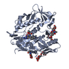 8fm3C  8fm4C 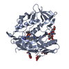 8fm7C  8fm8C C: citing same article ( |
|---|---|
| Similar structure data |
-Links
 PDBj
PDBj- Assembly
| Deposited unit | 
| ||||||||||||
|---|---|---|---|---|---|---|---|---|---|---|---|---|---|
| 1 | 
| ||||||||||||
| 2 | 
| ||||||||||||
| 3 | 
| ||||||||||||
| 4 | 
| ||||||||||||
| Unit cell |
|
-Components
| #1: Protein | Mass: 39755.664 Da / Num. of mol.: 4 Source method: isolated from a genetically manipulated source Source: (gene. exp.) HIV-1 06TG.HT008 (virus) / Production host:  Homo sapiens (human) Homo sapiens (human)#2: Sugar | ChemComp-NAG /   Type: D-saccharide, beta linking / Mass: 221.208 Da / Num. of mol.: 26 / Source method: obtained synthetically / Formula: C8H15NO6 Type: D-saccharide, beta linking / Mass: 221.208 Da / Num. of mol.: 26 / Source method: obtained synthetically / Formula: C8H15NO6#3: Chemical | ChemComp-Y2E / 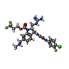  Mass: 587.954 Da / Num. of mol.: 4 / Source method: obtained synthetically / Formula: C24H26ClF4N7O4 / Feature type: SUBJECT OF INVESTIGATION Mass: 587.954 Da / Num. of mol.: 4 / Source method: obtained synthetically / Formula: C24H26ClF4N7O4 / Feature type: SUBJECT OF INVESTIGATION#4: Water | ChemComp-HOH / |  Mass: 18.015 Da / Num. of mol.: 209 / Source method: isolated from a natural source / Formula: H2O Mass: 18.015 Da / Num. of mol.: 209 / Source method: isolated from a natural source / Formula: H2OHas ligand of interest | Y | Has protein modification | Y | |
|---|
-Experimental details
-Experiment
| Experiment | Method: X-RAY DIFFRACTION / Number of used crystals: 1 |
|---|
- Sample preparation
| Crystal | Density Matthews: 2.67 Å3/Da / Density % sol: 53.94 % |
|---|---|
| Crystal grow | Temperature: 293 K / Method: vapor diffusion, hanging drop Details: 14% to 16% (w/v) PEG 1500, 0.1 M calcium chloride, 0.1 M imidazole pH 6.5 |
-Data collection
| Diffraction | Mean temperature: 100 K / Serial crystal experiment: N |
|---|---|
| Diffraction source | Source: SYNCHROTRON / Site: APS / Beamline: 24-ID-E / Wavelength: 0.9792 Å |
| Detector | Type: DECTRIS EIGER2 X 16M / Detector: PIXEL / Date: Nov 12, 2022 |
| Radiation | Protocol: SINGLE WAVELENGTH / Monochromatic (M) / Laue (L): M / Scattering type: x-ray |
| Radiation wavelength | Wavelength: 0.9792 Å / Relative weight: 1 |
| Reflection | Resolution: 1.88→48.81 Å / Num. obs: 88049 / % possible obs: 93.7 % / Redundancy: 11.9 % / Biso Wilson estimate: 30.52 Å2 / CC1/2: 0.997 / Rmerge(I) obs: 0.177 / Rpim(I) all: 0.052 / Net I/σ(I): 11.9 |
| Reflection shell | Resolution: 1.88→2.12 Å / Rmerge(I) obs: 1.666 / Num. unique obs: 4402 / CC1/2: 0.567 / Rpim(I) all: 0.514 |
- Processing
| Software |
| |||||||||||||||||||||||||||||||||||||||||||||||||||||||||||||||||||||||||||||||||||||||||||||||||||||||||
|---|---|---|---|---|---|---|---|---|---|---|---|---|---|---|---|---|---|---|---|---|---|---|---|---|---|---|---|---|---|---|---|---|---|---|---|---|---|---|---|---|---|---|---|---|---|---|---|---|---|---|---|---|---|---|---|---|---|---|---|---|---|---|---|---|---|---|---|---|---|---|---|---|---|---|---|---|---|---|---|---|---|---|---|---|---|---|---|---|---|---|---|---|---|---|---|---|---|---|---|---|---|---|---|---|---|---|
| Refinement | Method to determine structure: MOLECULAR REPLACEMENT / Resolution: 1.88→48.81 Å / SU ML: 0.2991 / Cross valid method: FREE R-VALUE / σ(F): 1.35 / Phase error: 39.6872 Stereochemistry target values: GeoStd + Monomer Library + CDL v1.2
| |||||||||||||||||||||||||||||||||||||||||||||||||||||||||||||||||||||||||||||||||||||||||||||||||||||||||
| Solvent computation | Shrinkage radii: 0.9 Å / VDW probe radii: 1.11 Å / Solvent model: FLAT BULK SOLVENT MODEL | |||||||||||||||||||||||||||||||||||||||||||||||||||||||||||||||||||||||||||||||||||||||||||||||||||||||||
| Displacement parameters | Biso mean: 50.13 Å2 | |||||||||||||||||||||||||||||||||||||||||||||||||||||||||||||||||||||||||||||||||||||||||||||||||||||||||
| Refinement step | Cycle: LAST / Resolution: 1.88→48.81 Å
| |||||||||||||||||||||||||||||||||||||||||||||||||||||||||||||||||||||||||||||||||||||||||||||||||||||||||
| Refine LS restraints |
| |||||||||||||||||||||||||||||||||||||||||||||||||||||||||||||||||||||||||||||||||||||||||||||||||||||||||
| LS refinement shell |
|
