National Institutes of Health/National Institute of General Medical Sciences (NIH/NIGMS)
5P41GM103533
United States
National Science Foundation (NSF, United States)
DGE-1762114
United States
Other private
Audacious Project philanthropic funds
National Institutes of Health/National Institute of General Medical Sciences (NIH/NIGMS)
R01 GM105691
United States
Other government
1S10D028581-01
Citation
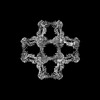
Journal: Nature / Year: 2025 Title: Four-component protein nanocages designed by programmed symmetry breaking. Authors: Sangmin Lee / Ryan D Kibler / Green Ahn / Yang Hsia / Andrew J Borst / Annika Philomin / Madison A Kennedy / Buwei Huang / Barry Stoddard / David Baker / Abstract: Four, eight or twenty C3 symmetric protein trimers can be arranged with tetrahedral, octahedral or icosahedral point group symmetry to generate closed cage-like structures. Viruses access more ...Four, eight or twenty C3 symmetric protein trimers can be arranged with tetrahedral, octahedral or icosahedral point group symmetry to generate closed cage-like structures. Viruses access more complex higher triangulation number icosahedral architectures by breaking perfect point group symmetry, but nature appears not to have explored similar symmetry breaking for tetrahedral or octahedral symmetries. Here we describe a general design strategy for building higher triangulation number architectures starting from regular polyhedra through pseudosymmetrization of trimeric building blocks. Electron microscopy confirms the structures of T = 4 cages with 48 (tetrahedral), 96 (octahedral) and 240 (icosahedral) subunits, each with 4 distinct chains and 6 different protein-protein interfaces, and diameters of 33 nm, 43 nm and 75 nm, respectively. Higher triangulation number viruses possess very sophisticated functionalities; our general route to higher triangulation number nanocages should similarly enable a next generation of multiple antigen-displaying vaccine candidates and targeted delivery vehicles.
History
Deposition
Dec 22, 2022
Deposition site: RCSB / Processing site: RCSB
Revision 1.0
Jun 28, 2023
Provider: repository / Type: Initial release
Revision 1.1
May 22, 2024
Group: Data collection / Category: chem_comp_atom / chem_comp_bond
Mass: 38677.871 Da / Num. of mol.: 1 Source method: isolated from a genetically manipulated source Source: (gene. exp.) synthetic construct (others) / Production host: Escherichia coli (E. coli)
Has protein modification
N
-
Experimental details
-
Experiment
Experiment
Method: X-RAY DIFFRACTION / Number of used crystals: 1
-
Sample preparation
Crystal
Density Matthews: 3.89 Å3/Da / Density % sol: 68.42 %
In the structure databanks used in Yorodumi, some data are registered as the other names, "COVID-19 virus" and "2019-nCoV". Here are the details of the virus and the list of structure data.
Jan 31, 2019. EMDB accession codes are about to change! (news from PDBe EMDB page)
EMDB accession codes are about to change! (news from PDBe EMDB page)
The allocation of 4 digits for EMDB accession codes will soon come to an end. Whilst these codes will remain in use, new EMDB accession codes will include an additional digit and will expand incrementally as the available range of codes is exhausted. The current 4-digit format prefixed with “EMD-” (i.e. EMD-XXXX) will advance to a 5-digit format (i.e. EMD-XXXXX), and so on. It is currently estimated that the 4-digit codes will be depleted around Spring 2019, at which point the 5-digit format will come into force.
The EM Navigator/Yorodumi systems omit the EMD- prefix.
Related info.:Q: What is EMD? / ID/Accession-code notation in Yorodumi/EM Navigator
Yorodumi is a browser for structure data from EMDB, PDB, SASBDB, etc.
This page is also the successor to EM Navigator detail page, and also detail information page/front-end page for Omokage search.
The word "yorodu" (or yorozu) is an old Japanese word meaning "ten thousand". "mi" (miru) is to see.
Related info.:EMDB / PDB / SASBDB / Comparison of 3 databanks / Yorodumi Search / Aug 31, 2016. New EM Navigator & Yorodumi / Yorodumi Papers / Jmol/JSmol / Function and homology information / Changes in new EM Navigator and Yorodumi
 Movie
Movie Controller

 Yorodumi
 Open data
 Basic information
 Components
 Keywords
 X-RAY DIFFRACTION /
  Authors
 United States, 6items
  Citation
 Structure visualization
 Molmil
 Downloads & links
 Other downloads
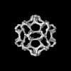


 PDBj
PDBj

 Assembly


 Sample preparation
 Processing