 Movie
Movie Controller

[English] 日本語
 Yorodumi
- PDB-8ffy: Cryo-electron microscopy structure of human mt-SerRS in complex w... -
+ Open data

- Basic information
| Entry | Database: PDB / ID: 8ffy | |||||||||||||||
|---|---|---|---|---|---|---|---|---|---|---|---|---|---|---|---|---|
| Title | Cryo-electron microscopy structure of human mt-SerRS in complex with mt-tRNA(UGA-TL) | |||||||||||||||
 Components Components |
| |||||||||||||||
 Keywords Keywords | LIGASE/RNA / tRNA / SerRS / transcription / ligase-RNA complex | |||||||||||||||
| Function / homology |  Function and homology information Function and homology informationmitochondrial seryl-tRNA aminoacylation / Mitochondrial tRNA aminoacylation / serine-tRNA ligase / serine-tRNA ligase activity / seryl-tRNA aminoacylation / tRNA binding / mitochondrial matrix / mitochondrion / RNA binding / ATP binding Similarity search - Function | |||||||||||||||
| Biological species |  Homo sapiens (human) Homo sapiens (human) | |||||||||||||||
| Method | ELECTRON MICROSCOPY / single particle reconstruction / cryo EM / Resolution: 3.6 Å | |||||||||||||||
 Authors Authors | Hirschi, M. / Kuhle, B. / Doerfel, L. / Schimmel, P. / Lander, G. | |||||||||||||||
| Funding support |  United States, 4items United States, 4items
| |||||||||||||||
 Citation Citation | Journal: Nat Commun / Year: 2023 Title: Structural basis for a degenerate tRNA identity code and the evolution of bimodal specificity in human mitochondrial tRNA recognition. Authors: Bernhard Kuhle / Marscha Hirschi / Lili K Doerfel / Gabriel C Lander / Paul Schimmel /  Abstract: Animal mitochondrial gene expression relies on specific interactions between nuclear-encoded aminoacyl-tRNA synthetases and mitochondria-encoded tRNAs. Their evolution involves an antagonistic ...Animal mitochondrial gene expression relies on specific interactions between nuclear-encoded aminoacyl-tRNA synthetases and mitochondria-encoded tRNAs. Their evolution involves an antagonistic interplay between strong mutation pressure on mtRNAs and selection pressure to maintain their essential function. To understand the molecular consequences of this interplay, we analyze the human mitochondrial serylation system, in which one synthetase charges two highly divergent mtRNA isoacceptors. We present the cryo-EM structure of human mSerRS in complex with mtRNA, and perform a structural and functional comparison with the mSerRS-mtRNA complex. We find that despite their common function, mtRNA and mtRNA show no constrain to converge on shared structural or sequence identity motifs for recognition by mSerRS. Instead, mSerRS evolved a bimodal readout mechanism, whereby a single protein surface recognizes degenerate identity features specific to each mtRNA. Our results show how the mutational erosion of mtRNAs drove a remarkable innovation of intermolecular specificity rules, with multiple evolutionary pathways leading to functionally equivalent outcomes. | |||||||||||||||
| History |
|
- Structure visualization
| Structure viewer | Molecule: MolmilJmol/JSmol |
|---|
- Downloads & links
-Download
| PDBx/mmCIF format | 8ffy.cif.gz | 201.6 KB | Display | PDBx/mmCIF format |
|---|---|---|---|---|
| PDB format | pdb8ffy.ent.gz | 153.2 KB | Display | PDB format |
| PDBx/mmJSON format | 8ffy.json.gz | Tree view | PDBx/mmJSON format | |
| Others |  Other downloads Other downloads |
-Validation report
| Arichive directory | https://data.pdbj.org/pub/pdb/validation_reports/ff/8ffyftp://data.pdbj.org/pub/pdb/validation_reports/ff/8ffy | HTTPS FTP |
|---|
-Related structure data
| Related structure data |  29070MC M: map data used to model this data C: citing same article ( |
|---|---|
| Similar structure data |
-Links
 PDBj
PDBj


- Assembly
| Deposited unit | 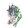
|
|---|---|
| 1 |
|
-Components
| #1: Protein | Mass: 58358.391 Da / Num. of mol.: 2 Source method: isolated from a genetically manipulated source Source: (gene. exp.) Homo sapiens (human) / Gene: SARS2, SARSM / Production host:  #2: RNA chain | | Mass: 21411.410 Da / Num. of mol.: 1 Source method: isolated from a genetically manipulated source Source: (gene. exp.) Homo sapiens (human) / Production host: #3: Chemical |   Mass: 433.397 Da / Num. of mol.: 2 / Source method: obtained synthetically / Formula: C13H19N7O8S Mass: 433.397 Da / Num. of mol.: 2 / Source method: obtained synthetically / Formula: C13H19N7O8SHas ligand of interest | N | |
|---|
-Experimental details
-Experiment
| Experiment | Method: ELECTRON MICROSCOPY |
|---|---|
| EM experiment | Aggregation state: PARTICLE / 3D reconstruction method: single particle reconstruction |
- Sample preparation
| Component | Name: Human mt-SerRS in complex with mt-tRNA(UGA-TL) and SerSA Type: COMPLEX / Entity ID: #1-#2 / Source: RECOMBINANT |
|---|---|
| Source (natural) | Organism: Homo sapiens (human) |
| Source (recombinant) | Organism: |
| Buffer solution | pH: 7.5 |
| Specimen | Embedding applied: NO / Shadowing applied: NO / Staining applied: NO / Vitrification applied: YES |
| Specimen support | Grid material: GOLD / Grid mesh size: 300 divisions/in. / Grid type: UltrAuFoil R1.2/1.3 |
| Vitrification | Cryogen name: ETHANE |
- Electron microscopy imaging
| Experimental equipment |  Model: Talos Arctica / Image courtesy: FEI Company |
|---|---|
| Microscopy | Model: FEI TALOS ARCTICA |
| Electron gun | Electron source:  FIELD EMISSION GUN / Accelerating voltage: 200 kV / Illumination mode: FLOOD BEAM FIELD EMISSION GUN / Accelerating voltage: 200 kV / Illumination mode: FLOOD BEAM |
| Electron lens | Mode: BRIGHT FIELD / Nominal defocus max: 1200 nm / Nominal defocus min: 800 nm |
| Image recording | Average exposure time: 11.8 sec. / Electron dose: 66 e/Å2 / Detector mode: COUNTING / Film or detector model: GATAN K2 SUMMIT (4k x 4k) / Num. of grids imaged: 1 / Num. of real images: 3448 |
- Processing
| EM software |
| ||||||||||||||||||||
|---|---|---|---|---|---|---|---|---|---|---|---|---|---|---|---|---|---|---|---|---|---|
| CTF correction | Type: PHASE FLIPPING AND AMPLITUDE CORRECTION | ||||||||||||||||||||
| Particle selection | Num. of particles selected: 7218136 | ||||||||||||||||||||
| Symmetry | Point symmetry: C1 (asymmetric) | ||||||||||||||||||||
| 3D reconstruction | Resolution: 3.6 Å / Resolution method: FSC 0.143 CUT-OFF / Num. of particles: 881655 / Symmetry type: POINT | ||||||||||||||||||||
| Atomic model building | Protocol: AB INITIO MODEL / Space: REAL |
