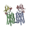
Mass: 39031.457 Da / Num. of mol.: 2 / Source method: isolated from a natural source / Source: (natural) Bos taurus (domestic cattle) / References: UniProt: P02699
#2: Antibody
NanobodyNb2
Mass: 13849.432 Da / Num. of mol.: 2 Source method: isolated from a genetically manipulated source Source: (gene. exp.) Lama glama (llama) / Production host: Escherichia coli (E. coli)
Method: X-RAY DIFFRACTION / Number of used crystals: 1
-
Sample preparation
Crystal
Density Matthews: 4.93 Å3/Da / Density % sol: 75.03 %
Crystal grow
Temperature: 277.15 K / Method: vapor diffusion, sitting drop / pH: 8 Details: 30% PEG 600 0.1 M Tricine pH 8 1.2 mM F3215-0002 2% IPTG 50 mM phenol 5 mM EDTA 5 mM beta mercaptoethanol 50 mM Cyglu-4
-
Data collection
Diffraction
Mean temperature: 100 K / Serial crystal experiment: N
Protocol: SINGLE WAVELENGTH / Monochromatic (M) / Laue (L): M / Scattering type: x-ray
Radiation wavelength
Wavelength: 0.97918 Å / Relative weight: 1
Reflection
Resolution: 3.7→50 Å / Num. obs: 20570 / % possible obs: 99.8 % / Redundancy: 9.8 % / CC1/2: 0.999 / Rmerge(I) obs: 0.094 / Net I/σ(I): 10.22
Reflection shell
Resolution: 3.7→3.92 Å / Rmerge(I) obs: 2.831 / Mean I/σ(I) obs: 0.71 / Num. unique obs: 3236 / CC1/2: 0.378
-
Processing
Software
Name
Version
Classification
REFMAC
5.8.0352
refinement
PDB_EXTRACT
3.27
dataextraction
XDS
datareduction
XDS
datascaling
PHASER
phasing
Refinement
Method to determine structure: MOLECULAR REPLACEMENT / Resolution: 3.7→49.76 Å / Cor.coef. Fo:Fc: 0.874 / Cor.coef. Fo:Fc free: 0.862 / SU B: 55.867 / SU ML: 0.718 / Cross valid method: THROUGHOUT / σ(F): 0 / ESU R Free: 0.741 / Stereochemistry target values: MAXIMUM LIKELIHOOD Details: HYDROGENS HAVE BEEN ADDED IN THE RIDING POSITIONS U VALUES : REFINED INDIVIDUALLY
Rfactor
Num. reflection
% reflection
Selection details
Rfree
0.3318
1029
5 %
RANDOM
Rwork
0.2809
-
-
-
obs
0.2834
19540
99.82 %
-
Solvent computation
Ion probe radii: 0.9 Å / Shrinkage radii: 0.9 Å / VDW probe radii: 1.3 Å / Solvent model: MASK
In the structure databanks used in Yorodumi, some data are registered as the other names, "COVID-19 virus" and "2019-nCoV". Here are the details of the virus and the list of structure data.
Jan 31, 2019. EMDB accession codes are about to change! (news from PDBe EMDB page)
EMDB accession codes are about to change! (news from PDBe EMDB page)
The allocation of 4 digits for EMDB accession codes will soon come to an end. Whilst these codes will remain in use, new EMDB accession codes will include an additional digit and will expand incrementally as the available range of codes is exhausted. The current 4-digit format prefixed with “EMD-” (i.e. EMD-XXXX) will advance to a 5-digit format (i.e. EMD-XXXXX), and so on. It is currently estimated that the 4-digit codes will be depleted around Spring 2019, at which point the 5-digit format will come into force.
The EM Navigator/Yorodumi systems omit the EMD- prefix.
Related info.:Q: What is EMD? / ID/Accession-code notation in Yorodumi/EM Navigator
Yorodumi is a browser for structure data from EMDB, PDB, SASBDB, etc.
This page is also the successor to EM Navigator detail page, and also detail information page/front-end page for Omokage search.
The word "yorodu" (or yorozu) is an old Japanese word meaning "ten thousand". "mi" (miru) is to see.
Related info.:EMDB / PDB / SASBDB / Comparison of 3 databanks / Yorodumi Search / Aug 31, 2016. New EM Navigator & Yorodumi / Yorodumi Papers / Jmol/JSmol / Function and homology information / Changes in new EM Navigator and Yorodumi
 Movie
Movie Controller

 Yorodumi
 Open data
 Basic information
 Components
 Keywords
 Function and homology information
 X-RAY DIFFRACTION /
  Authors
 United States, 3items
  Citation
 Structure visualization
 Downloads & links
 Other downloads
 PDBj
PDBj


 Assembly
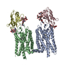


 Mass: 284.436 Da / Num. of mol.: 2 / Source method: obtained synthetically / Formula: C20H28O / Feature type: SUBJECT OF INVESTIGATION
 Sample preparation
 Processing