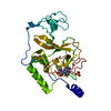
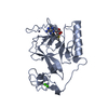
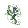
登録情報 データベース : PDB / ID : 8fbgタイトル Crystal structure of NSD1 Mutant-Y1869C Histone-lysine N-methyltransferase, H3 lysine-36 specific キーワード / / / / 機能・相同性 分子機能 ドメイン・相同性 構成要素
/ / / / / / / / / / / / / / / / / / / / / / / / / / / / / / / / / / / / / / / / / / / / / / / / / / / / / / / / / / / / / / / / / / / / / / / / / / / / / 生物種 Mus musculus (ハツカネズミ)手法 / / 解像度 : 2.29 Å データ登録者 Providokhina, K. / Dong, A. / Arrowsmith, C.H. / Edwards, A.M. / Min, J. / Structural Genomics Consortium (SGC) 資金援助 1件 ジャーナル : To Be Published タイトル : Crystal structure of NSD1著者 : Providokhina, K. / Dong, A. / Arrowsmith, C.H. / Edwards, A.M. / Min, J. / Structural Genomics Consortium (SGC) 履歴 登録 2022年11月29日 登録サイト / 処理サイト 改定 1.0 2023年1月18日 Provider / タイプ 改定 1.1 2023年10月25日 Group / Refinement descriptionカテゴリ / chem_comp_bond / pdbx_initial_refinement_model
すべて表示 表示を減らす
 ムービー
ムービー コントローラー


 データを開く
 基本情報
基本情報 要素
要素 キーワード
キーワード 機能・相同性情報
 X線回折 /
  データ登録者
 引用
引用 構造の表示
構造の表示 ダウンロードとリンク
 その他のダウンロード
 PDBj
PDBj
 集合体
集合体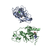


 分子量: 65.409 Da / 分子数: 7 / 由来タイプ: 合成 / 式: Zn
 分子量: 40.078 Da / 分子数: 4 / 由来タイプ: 合成 / 式: Ca
 分子量: 398.437 Da / 分子数: 2 / 由来タイプ: 合成 / 式: C15H22N6O5S
 分子量: 18.015 Da / 分子数: 170 / 由来タイプ: 天然 / 式: H2O
 試料調製
試料調製 解析
解析