 Movie
Movie Controller

[English] 日本語
 Yorodumi
- PDB-8f73: Crystal structure of Pseudomonas aeruginosa UDP-glucose phosphory... -
+ Open data

- Basic information
| Entry | Database: PDB / ID: 8f73 | ||||||
|---|---|---|---|---|---|---|---|
| Title | Crystal structure of Pseudomonas aeruginosa UDP-glucose phosphorylase in complex with UDP-glucose | ||||||
 Components Components | UTP--glucose-1-phosphate uridylyltransferase | ||||||
 Keywords Keywords | TRANSFERASE / UGP / UDP-Glucose pyrophosphorylase / UTP:glucose-1-phosphate uridylyltransferase | ||||||
| Function / homology |  Function and homology information Function and homology informationUTP-glucose-1-phosphate uridylyltransferase / UTP:glucose-1-phosphate uridylyltransferase activity / UDP-alpha-D-glucose metabolic process / lipopolysaccharide core region biosynthetic process Similarity search - Function | ||||||
| Biological species |  Pseudomonas aeruginosa PAO1 (bacteria) Pseudomonas aeruginosa PAO1 (bacteria) | ||||||
| Method |  X-RAY DIFFRACTION / SYNCHROTRON / MOLECULAR REPLACEMENT / Resolution: 2.9 Å X-RAY DIFFRACTION / SYNCHROTRON / MOLECULAR REPLACEMENT / Resolution: 2.9 Å | ||||||
 Authors Authors | Dirr, L. / Fuehring, J. | ||||||
| Funding support |  Australia, 1items Australia, 1items
| ||||||
 Citation Citation | Journal: Mbio / Year: 2024 Title: Tetramerization is essential for the enzymatic function of the Pseudomonas aeruginosa virulence factor UDP-glucose pyrophosphorylase. Authors: Dirr, L. / Cleeves, S. / Ramon Roth, I. / Li, L. / Fiebig, T. / Ve, T. / Haussler, S. / Braun, A. / von Itzstein, M. / Fuhring, J.I. | ||||||
| History |
|
- Structure visualization
| Structure viewer | Molecule: MolmilJmol/JSmol |
|---|
- Downloads & links
-Download
| PDBx/mmCIF format | 8f73.cif.gz | 888 KB | Display | PDBx/mmCIF format |
|---|---|---|---|---|
| PDB format | pdb8f73.ent.gz | 690.3 KB | Display | PDB format |
| PDBx/mmJSON format | 8f73.json.gz | Tree view | PDBx/mmJSON format | |
| Others |  Other downloads Other downloads |
-Validation report
| Summary document | 8f73_validation.pdf.gz | 3 MB | Display | wwPDB validaton report |
|---|---|---|---|---|
| Full document | 8f73_full_validation.pdf.gz | 3.1 MB | Display | |
| Data in XML | 8f73_validation.xml.gz | 80.7 KB | Display | |
| Data in CIF | 8f73_validation.cif.gz | 105.8 KB | Display | |
| Arichive directory | https://data.pdbj.org/pub/pdb/validation_reports/f7/8f73ftp://data.pdbj.org/pub/pdb/validation_reports/f7/8f73 | HTTPS FTP |
-Related structure data
| Related structure data | 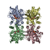 3jukS S: Starting model for refinement |
|---|---|
| Similar structure data |
-Links
 PDBj
PDBj

- Assembly
| Deposited unit | 
| ||||||||||||
|---|---|---|---|---|---|---|---|---|---|---|---|---|---|
| 1 | 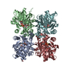
| ||||||||||||
| 2 | 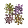
| ||||||||||||
| Unit cell |
|
-Components
-Protein , 1 types, 8 molecules ABCDEFGH
| #1: Protein | Mass: 31824.680 Da / Num. of mol.: 8 Source method: isolated from a genetically manipulated source Source: (gene. exp.) Pseudomonas aeruginosa PAO1 (bacteria) / Gene: galU, PA2023 / Production host: References: UniProt: Q9I291, UTP-glucose-1-phosphate uridylyltransferase |
|---|
-Non-polymers , 5 types, 122 molecules 


| #2: Chemical | ChemComp-UPG /  Mass: 566.302 Da / Num. of mol.: 8 / Source method: obtained synthetically / Formula: C15H24N2O17P2 / Feature type: SUBJECT OF INVESTIGATION Mass: 566.302 Da / Num. of mol.: 8 / Source method: obtained synthetically / Formula: C15H24N2O17P2 / Feature type: SUBJECT OF INVESTIGATION#3: Chemical |  Mass: 62.068 Da / Num. of mol.: 3 / Source method: obtained synthetically / Formula: C2H6O2 Mass: 62.068 Da / Num. of mol.: 3 / Source method: obtained synthetically / Formula: C2H6O2#4: Chemical | ChemComp-MG /  Mass: 24.305 Da / Num. of mol.: 8 / Source method: obtained synthetically / Formula: Mg Mass: 24.305 Da / Num. of mol.: 8 / Source method: obtained synthetically / Formula: Mg#5: Chemical | ChemComp-ACT / |  Mass: 59.044 Da / Num. of mol.: 1 / Source method: obtained synthetically / Formula: C2H3O2 Mass: 59.044 Da / Num. of mol.: 1 / Source method: obtained synthetically / Formula: C2H3O2#6: Water | ChemComp-HOH / | Mass: 18.015 Da / Num. of mol.: 102 / Source method: isolated from a natural source / Formula: H2O |
|---|
-Details
| Has ligand of interest | Y |
|---|---|
| Has protein modification | Y |
-Experimental details
-Experiment
| Experiment | Method: X-RAY DIFFRACTION / Number of used crystals: 1 |
|---|
- Sample preparation
| Crystal | Density Matthews: 2.95 Å3/Da / Density % sol: 58.33 % |
|---|---|
| Crystal grow | Temperature: 293 K / Method: vapor diffusion, hanging drop Details: 0.1 M sodium acetate trihydrate pH 4.6 2.0 M sodium formate |
-Data collection
| Diffraction | Mean temperature: 100 K / Serial crystal experiment: N |
|---|---|
| Diffraction source | Source: SYNCHROTRON / Site: Australian Synchrotron / Beamline: MX2 / Wavelength: 0.953722 Å |
| Detector | Type: DECTRIS EIGER X 16M / Detector: PIXEL / Date: Mar 3, 2019 |
| Radiation | Protocol: SINGLE WAVELENGTH / Monochromatic (M) / Laue (L): M / Scattering type: x-ray |
| Radiation wavelength | Wavelength: 0.953722 Å / Relative weight: 1 |
| Reflection | Resolution: 2.9→48.52 Å / Num. obs: 65768 / % possible obs: 99.7 % / Redundancy: 3.9 % / Biso Wilson estimate: 60.43 Å2 / CC1/2: 0.96 / Rmerge(I) obs: 0.109 / Net I/σ(I): 9.3 |
| Reflection shell | Resolution: 2.9→2.97 Å / Rmerge(I) obs: 0.86 / Mean I/σ(I) obs: 1.8 / Num. unique obs: 4608 / CC1/2: 1 / Rpim(I) all: 0.58 |
- Processing
| Software |
| ||||||||||||||||||||||||||||||||||||||||||||||||||||||||||||||||||||||||||||||||||||||||||||||||||||||||||||||||||||||||||||||||||||||||||||||||||||||||||||||||||||||||
|---|---|---|---|---|---|---|---|---|---|---|---|---|---|---|---|---|---|---|---|---|---|---|---|---|---|---|---|---|---|---|---|---|---|---|---|---|---|---|---|---|---|---|---|---|---|---|---|---|---|---|---|---|---|---|---|---|---|---|---|---|---|---|---|---|---|---|---|---|---|---|---|---|---|---|---|---|---|---|---|---|---|---|---|---|---|---|---|---|---|---|---|---|---|---|---|---|---|---|---|---|---|---|---|---|---|---|---|---|---|---|---|---|---|---|---|---|---|---|---|---|---|---|---|---|---|---|---|---|---|---|---|---|---|---|---|---|---|---|---|---|---|---|---|---|---|---|---|---|---|---|---|---|---|---|---|---|---|---|---|---|---|---|---|---|---|---|---|---|---|
| Refinement | Method to determine structure: MOLECULAR REPLACEMENT Starting model: 3JUK Resolution: 2.9→46.98 Å / SU ML: 0.3435 / Cross valid method: FREE R-VALUE / σ(F): 1.36 / Phase error: 25.8171 Stereochemistry target values: GeoStd + Monomer Library + CDL v1.2
| ||||||||||||||||||||||||||||||||||||||||||||||||||||||||||||||||||||||||||||||||||||||||||||||||||||||||||||||||||||||||||||||||||||||||||||||||||||||||||||||||||||||||
| Solvent computation | Shrinkage radii: 0.9 Å / VDW probe radii: 1.11 Å / Solvent model: FLAT BULK SOLVENT MODEL | ||||||||||||||||||||||||||||||||||||||||||||||||||||||||||||||||||||||||||||||||||||||||||||||||||||||||||||||||||||||||||||||||||||||||||||||||||||||||||||||||||||||||
| Displacement parameters | Biso mean: 65.16 Å2 | ||||||||||||||||||||||||||||||||||||||||||||||||||||||||||||||||||||||||||||||||||||||||||||||||||||||||||||||||||||||||||||||||||||||||||||||||||||||||||||||||||||||||
| Refinement step | Cycle: LAST / Resolution: 2.9→46.98 Å
| ||||||||||||||||||||||||||||||||||||||||||||||||||||||||||||||||||||||||||||||||||||||||||||||||||||||||||||||||||||||||||||||||||||||||||||||||||||||||||||||||||||||||
| Refine LS restraints |
| ||||||||||||||||||||||||||||||||||||||||||||||||||||||||||||||||||||||||||||||||||||||||||||||||||||||||||||||||||||||||||||||||||||||||||||||||||||||||||||||||||||||||
| LS refinement shell |
|
