 Movie
Movie Controller

[English] 日本語
 Yorodumi
- PDB-8f43: HNH Nuclease Domain from G. stearothermophilus Cas9, K597A mutant -
+ Open data

- Basic information
| Entry | Database: PDB / ID: 8f43 | ||||||
|---|---|---|---|---|---|---|---|
| Title | HNH Nuclease Domain from G. stearothermophilus Cas9, K597A mutant | ||||||
 Components Components | CRISPR-associated endonuclease Cas9 | ||||||
 Keywords Keywords | HYDROLASE / nuclease domain / CRISPR Cas9 / DNA binding protein | ||||||
| Function / homology |  Function and homology information Function and homology informationmaintenance of CRISPR repeat elements / endonuclease activity / defense response to virus / Hydrolases; Acting on ester bonds / DNA binding / RNA binding / metal ion binding Similarity search - Function | ||||||
| Biological species |   Geobacillus stearothermophilus (bacteria) Geobacillus stearothermophilus (bacteria) | ||||||
| Method |  X-RAY DIFFRACTION / SYNCHROTRON / MOLECULAR REPLACEMENT / Resolution: 1.37 Å X-RAY DIFFRACTION / SYNCHROTRON / MOLECULAR REPLACEMENT / Resolution: 1.37 Å | ||||||
 Authors Authors | D'Ordine, A.M. / Belato, H.B. / Lisi, G.P. / Jogl, G. | ||||||
| Funding support |  United States, 1items United States, 1items
| ||||||
 Citation Citation | Journal: J.Chem.Phys. / Year: 2022 Title: Disruption of electrostatic contacts in the HNH nuclease from a thermophilic Cas9 rewires allosteric motions and enhances high-temperature DNA cleavage. Authors: Belato, H.B. / Norbrun, C. / Luo, J. / Pindi, C. / Sinha, S. / D'Ordine, A.M. / Jogl, G. / Palermo, G. / Lisi, G.P. #1: Journal: Acta Crystallogr., Sect. D: Biol. Crystallogr. / Year: 2012Title: Towards automated crystallographic structure refinement with phenix.refine. Authors: Afonine, P.V. / Grosse-Kunstleve, R.W. / Echols, N. / Headd, J.J. / Moriarty, N.W. / Mustyakimov, M. / Terwilliger, T.C. / Urzhumtsev, A. / Zwart, P.H. / Adams, P.D. | ||||||
| History |
|
- Structure visualization
| Structure viewer | Molecule: MolmilJmol/JSmol |
|---|
- Downloads & links
-Download
| PDBx/mmCIF format | 8f43.cif.gz | 73.6 KB | Display | PDBx/mmCIF format |
|---|---|---|---|---|
| PDB format | pdb8f43.ent.gz | 45.3 KB | Display | PDB format |
| PDBx/mmJSON format | 8f43.json.gz | Tree view | PDBx/mmJSON format | |
| Others |  Other downloads Other downloads |
-Validation report
| Arichive directory | https://data.pdbj.org/pub/pdb/validation_reports/f4/8f43ftp://data.pdbj.org/pub/pdb/validation_reports/f4/8f43 | HTTPS FTP |
|---|
-Related structure data
| Related structure data | 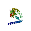 7mpzS S: Starting model for refinement |
|---|---|
| Similar structure data |
-Links
 PDBj
PDBj
- Assembly
| Deposited unit | 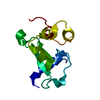
| ||||||||||||
|---|---|---|---|---|---|---|---|---|---|---|---|---|---|
| 1 |
| ||||||||||||
| Unit cell |
| ||||||||||||
| Components on special symmetry positions |
|
-Components
| #1: Protein | Mass: 13020.691 Da / Num. of mol.: 1 / Fragment: Nuclease Domain / Mutation: K597A Source method: isolated from a genetically manipulated source Source: (gene. exp.) Geobacillus stearothermophilus (bacteria)Gene: cas9, GS458_0313 / Production host: References: UniProt: A0A250DVH8, Hydrolases; Acting on ester bonds |
|---|---|
| #2: Water | ChemComp-HOH /  Mass: 18.015 Da / Num. of mol.: 113 / Source method: isolated from a natural source / Formula: H2O Mass: 18.015 Da / Num. of mol.: 113 / Source method: isolated from a natural source / Formula: H2O |
-Experimental details
-Experiment
| Experiment | Method: X-RAY DIFFRACTION / Number of used crystals: 1 |
|---|
- Sample preparation
| Crystal | Density Matthews: 2.01 Å3/Da / Density % sol: 38.9 % |
|---|---|
| Crystal grow | Temperature: 298 K / Method: vapor diffusion, sitting drop Details: 0.2M calcium chloride hexahydrate, 0.1M HEPES pH 7.0, 20% polyethylene glycol 6000 |
-Data collection
| Diffraction | Mean temperature: 100 K / Serial crystal experiment: N |
|---|---|
| Diffraction source | Source: SYNCHROTRON / Site: NSLS-II / Beamline: 17-ID-1 / Wavelength: 0.920105 Å |
| Detector | Type: DECTRIS EIGER X 9M / Detector: PIXEL / Date: Jul 18, 2022 |
| Radiation | Protocol: SINGLE WAVELENGTH / Monochromatic (M) / Laue (L): M / Scattering type: x-ray |
| Radiation wavelength | Wavelength: 0.920105 Å / Relative weight: 1 |
| Reflection | Resolution: 1.37→28.58 Å / Num. obs: 22933 / % possible obs: 100 % / Redundancy: 13.1 % / Biso Wilson estimate: 11.83 Å2 / CC1/2: 0.988 / Net I/σ(I): 7.3 |
| Reflection shell | Resolution: 1.37→1.39 Å / Num. unique obs: 1146 / CC1/2: 0.878 |
- Processing
| Software |
| |||||||||||||||||||||||||||||||||||||||||||||||||||||||||||||||
|---|---|---|---|---|---|---|---|---|---|---|---|---|---|---|---|---|---|---|---|---|---|---|---|---|---|---|---|---|---|---|---|---|---|---|---|---|---|---|---|---|---|---|---|---|---|---|---|---|---|---|---|---|---|---|---|---|---|---|---|---|---|---|---|---|
| Refinement | Method to determine structure: MOLECULAR REPLACEMENT Starting model: 7MPZ Resolution: 1.37→26.92 Å / Cross valid method: FREE R-VALUE
| |||||||||||||||||||||||||||||||||||||||||||||||||||||||||||||||
| Refinement step | Cycle: LAST / Resolution: 1.37→26.92 Å
| |||||||||||||||||||||||||||||||||||||||||||||||||||||||||||||||
| Refine LS restraints |
| |||||||||||||||||||||||||||||||||||||||||||||||||||||||||||||||
| LS refinement shell |
| |||||||||||||||||||||||||||||||||||||||||||||||||||||||||||||||
| Refinement TLS params. | Method: refined / Origin x: 14.6869403231 Å / Origin y: 13.3420374967 Å / Origin z: 4.73755081448 Å
| |||||||||||||||||||||||||||||||||||||||||||||||||||||||||||||||
| Refinement TLS group | Selection details: all |
