 Movie
Movie Controller


+ Open data

- Basic information
| Entry | Database: PDB / ID: 7mpz | |||||||||
|---|---|---|---|---|---|---|---|---|---|---|
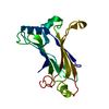
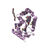
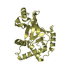
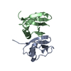
| Title | HNH Nuclease Domain from G. stearothermophilus Cas9 | |||||||||
 Components Components | CRISPR-associated endonuclease Cas9 | |||||||||
 Keywords Keywords | DNA BINDING PROTEIN / nuclease domain / CRISPR Cas9 | |||||||||
| Function / homology |  Function and homology information Function and homology informationmaintenance of CRISPR repeat elements / endonuclease activity / defense response to virus / Hydrolases; Acting on ester bonds / DNA binding / RNA binding / metal ion binding Similarity search - Function | |||||||||
| Biological species |   Geobacillus stearothermophilus (bacteria) Geobacillus stearothermophilus (bacteria) | |||||||||
| Method |  X-RAY DIFFRACTION / MOLECULAR REPLACEMENT / Resolution: 2.04 Å X-RAY DIFFRACTION / MOLECULAR REPLACEMENT / Resolution: 2.04 Å | |||||||||
 Authors Authors | D'Ordine, A.M. / Belato, H.B. / Lisi, G.P. / Jogl, G. | |||||||||
| Funding support |  United States, 2items United States, 2items
| |||||||||
 Citation Citation | Journal: J.Struct.Biol. / Year: 2021 Title: Structural and dynamic insights into the HNH nuclease of divergent Cas9 species. Authors: Belato, H.B. / D'Ordine, A.M. / Nierzwicki, L. / Arantes, P.R. / Jogl, G. / Palermo, G. / Lisi, G.P. | |||||||||
| History |
|
- Structure visualization
| Structure viewer | Molecule: MolmilJmol/JSmol |
|---|
- Downloads & links
-Download
| PDBx/mmCIF format | 7mpz.cif.gz | 87.3 KB | Display | PDBx/mmCIF format |
|---|---|---|---|---|
| PDB format | pdb7mpz.ent.gz | 53.9 KB | Display | PDB format |
| PDBx/mmJSON format | 7mpz.json.gz | Tree view | PDBx/mmJSON format | |
| Others |  Other downloads Other downloads |
-Validation report
| Arichive directory | https://data.pdbj.org/pub/pdb/validation_reports/mp/7mpzftp://data.pdbj.org/pub/pdb/validation_reports/mp/7mpz | HTTPS FTP |
|---|
-Related structure data
| Related structure data | 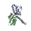 6j9nS S: Starting model for refinement |
|---|---|
| Similar structure data |


-Links
 PDBj
PDBj
- Assembly
| Deposited unit | 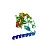
| ||||||||||||
|---|---|---|---|---|---|---|---|---|---|---|---|---|---|
| 1 |
| ||||||||||||
| Unit cell |
|
-Components
| #1: Protein | Mass: 18507.867 Da / Num. of mol.: 1 Source method: isolated from a genetically manipulated source Source: (gene. exp.) Geobacillus stearothermophilus (bacteria)Gene: cas9, GS458_0313 / Production host: References: UniProt: A0A250DVH8, Hydrolases; Acting on ester bonds |
|---|---|
| #2: Water | ChemComp-HOH /  Mass: 18.015 Da / Num. of mol.: 67 / Source method: isolated from a natural source / Formula: H2O Mass: 18.015 Da / Num. of mol.: 67 / Source method: isolated from a natural source / Formula: H2O |
-Experimental details
-Experiment
| Experiment | Method: X-RAY DIFFRACTION / Number of used crystals: 1 |
|---|
- Sample preparation
| Crystal | Density Matthews: 2.71 Å3/Da / Density % sol: 54.59 % |
|---|---|
| Crystal grow | Temperature: 298 K / Method: vapor diffusion, sitting drop Details: 0.12 M [0.3 M diethylene glycol, 0.3 M triethylene glycol, 0.3 M tetraethylene glycol, 0.3 M pentaethylene glycol], 0.1 M [MES and imidazole] pH 6.5, 30% (v/v) [40% (v/v) glycerol, 20% (w/v) ...Details: 0.12 M [0.3 M diethylene glycol, 0.3 M triethylene glycol, 0.3 M tetraethylene glycol, 0.3 M pentaethylene glycol], 0.1 M [MES and imidazole] pH 6.5, 30% (v/v) [40% (v/v) glycerol, 20% (w/v) polyethylene glycol 4000] |
-Data collection
| Diffraction | Mean temperature: 100 K / Serial crystal experiment: N |
|---|---|
| Diffraction source | Source: SEALED TUBE / Type: RIGAKU MICROMAX-003 / Wavelength: 1.54178 Å |
| Detector | Type: RIGAKU SATURN 944 / Detector: CCD / Date: Sep 5, 2019 |
| Radiation | Protocol: SINGLE WAVELENGTH / Monochromatic (M) / Laue (L): M / Scattering type: x-ray |
| Radiation wavelength | Wavelength: 1.54178 Å / Relative weight: 1 |
| Reflection | Resolution: 2.04→38.76 Å / Num. obs: 24738 / % possible obs: 99.85 % / Redundancy: 7.3 % / Biso Wilson estimate: 39.68 Å2 / CC1/2: 1 / CC star: 1 / Net I/σ(I): 20.31 |
| Reflection shell | Resolution: 2.04→2.11 Å / Redundancy: 5.6 % / Num. unique obs: 2458 / CC1/2: 0.539 / CC star: 0.837 / % possible all: 99.39 |
- Processing
| Software |
| ||||||||||||||||||||||||||||||||||||||||||||||||||||||||||||||||||||||
|---|---|---|---|---|---|---|---|---|---|---|---|---|---|---|---|---|---|---|---|---|---|---|---|---|---|---|---|---|---|---|---|---|---|---|---|---|---|---|---|---|---|---|---|---|---|---|---|---|---|---|---|---|---|---|---|---|---|---|---|---|---|---|---|---|---|---|---|---|---|---|---|
| Refinement | Method to determine structure: MOLECULAR REPLACEMENT Starting model: 6J9N Resolution: 2.04→38.76 Å / SU ML: 0.2174 / Cross valid method: FREE R-VALUE / σ(F): 1.34 / Phase error: 23.2523 Stereochemistry target values: GeoStd + Monomer Library + CDL v1.2
| ||||||||||||||||||||||||||||||||||||||||||||||||||||||||||||||||||||||
| Solvent computation | Shrinkage radii: 0.9 Å / VDW probe radii: 1.11 Å / Solvent model: FLAT BULK SOLVENT MODEL | ||||||||||||||||||||||||||||||||||||||||||||||||||||||||||||||||||||||
| Displacement parameters | Biso mean: 49.48 Å2 | ||||||||||||||||||||||||||||||||||||||||||||||||||||||||||||||||||||||
| Refinement step | Cycle: LAST / Resolution: 2.04→38.76 Å
| ||||||||||||||||||||||||||||||||||||||||||||||||||||||||||||||||||||||
| Refine LS restraints |
| ||||||||||||||||||||||||||||||||||||||||||||||||||||||||||||||||||||||
| LS refinement shell |
| ||||||||||||||||||||||||||||||||||||||||||||||||||||||||||||||||||||||
| Refinement TLS params. | Method: refined / Origin x: -16.5146904761 Å / Origin y: -0.401937909455 Å / Origin z: -3.73874303122 Å
| ||||||||||||||||||||||||||||||||||||||||||||||||||||||||||||||||||||||
| Refinement TLS group | Selection details: all |
