 Movie
Movie Controller


+ Open data

- Basic information
| Entry | Database: PDB / ID: 8f0v | ||||||
|---|---|---|---|---|---|---|---|
| Title | Lipocalin-like Milk protein-2 - E38A mutant | ||||||
 Components Components | Milk protein | ||||||
 Keywords Keywords | LIPID BINDING PROTEIN / Lipocalin / Mutation / cockroach milk / Lili Mip. | ||||||
| Function / homology | Calycin / metal ion binding / Milk protein Function and homology information Function and homology information | ||||||
| Biological species |  Diploptera punctata (Pacific beetle cockroach) Diploptera punctata (Pacific beetle cockroach) | ||||||
| Method |  X-RAY DIFFRACTION / SYNCHROTRON / MOLECULAR REPLACEMENT / Resolution: 2.951 Å X-RAY DIFFRACTION / SYNCHROTRON / MOLECULAR REPLACEMENT / Resolution: 2.951 Å | ||||||
 Authors Authors | Subramanian, R. / KanagaVijayan, D. | ||||||
| Funding support | 1items
| ||||||
 Citation Citation | Journal: Plos One / Year: 2023 Title: Variability in phenylalanine side chain conformations facilitates broad substrate tolerance of fatty acid binding in cockroach milk proteins. Authors: Santhakumari, P.R. / Dhanabalan, K. / Virani, S. / Hopf-Jannasch, A.S. / Benoit, J.B. / Chopra, G. / Subramanian, R. #1: Journal: Biochim Biophys Acta Gen Subj / Year: 2022Title: Structure of recombinantly expressed cockroach Lili-Mip protein in glycosylated and deglycosylated forms. Authors: KanagaVijayan, D. / Subramanian, R. / Santhakumari, P.R. / Chavas, L.M.G. / Subramanian, R. / Banerjee, S. #2: Journal: IUCrJ / Year: 2016Title: Structure of a heterogeneous, glycosylated, lipid-bound, in vivo-grown protein crystal at atomic resolution from the viviparous cockroach Diploptera punctata. Authors: Banerjee, S. / Coussens, N.P. / Gallat, F.X. / Sathyanarayanan, N. / Srikanth, J. / Yagi, K.J. / Gray, J.S. / Tobe, S.S. / Stay, B. / Chavas, L.M. / Ramaswamy, S. | ||||||
| History |
|
- Structure visualization
| Structure viewer | Molecule: MolmilJmol/JSmol |
|---|
- Downloads & links
-Download
| PDBx/mmCIF format | 8f0v.cif.gz | 78.1 KB | Display | PDBx/mmCIF format |
|---|---|---|---|---|
| PDB format | pdb8f0v.ent.gz | 56.6 KB | Display | PDB format |
| PDBx/mmJSON format | 8f0v.json.gz | Tree view | PDBx/mmJSON format | |
| Others |  Other downloads Other downloads |
-Validation report
| Summary document | 8f0v_validation.pdf.gz | 448 KB | Display | wwPDB validaton report |
|---|---|---|---|---|
| Full document | 8f0v_full_validation.pdf.gz | 450 KB | Display | |
| Data in XML | 8f0v_validation.xml.gz | 8.8 KB | Display | |
| Data in CIF | 8f0v_validation.cif.gz | 10.6 KB | Display | |
| Arichive directory | https://data.pdbj.org/pub/pdb/validation_reports/f0/8f0vftp://data.pdbj.org/pub/pdb/validation_reports/f0/8f0v | HTTPS FTP |
-Related structure data
| Related structure data | 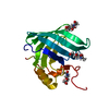 8f0yC  7bkxS S: Starting model for refinement C: citing same article ( |
|---|---|
| Similar structure data |
-Links
 PDBj
PDBj

- Assembly
| Deposited unit | 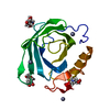
| ||||||||
|---|---|---|---|---|---|---|---|---|---|
| 1 |
| ||||||||
| Unit cell |
| ||||||||
| Components on special symmetry positions |
|
-Components
| #1: Protein | Mass: 17958.211 Da / Num. of mol.: 1 / Mutation: E38A Source method: isolated from a genetically manipulated source Source: (gene. exp.) Diploptera punctata (Pacific beetle cockroach)Production host:  | ||||||||
|---|---|---|---|---|---|---|---|---|---|
| #2: Sugar |   Type: D-saccharide, beta linking / Mass: 221.208 Da / Num. of mol.: 3 / Source method: obtained synthetically / Formula: C8H15NO6 Type: D-saccharide, beta linking / Mass: 221.208 Da / Num. of mol.: 3 / Source method: obtained synthetically / Formula: C8H15NO6#3: Chemical |   Mass: 65.409 Da / Num. of mol.: 3 / Source method: obtained synthetically / Formula: Zn Mass: 65.409 Da / Num. of mol.: 3 / Source method: obtained synthetically / Formula: Zn#4: Water | ChemComp-HOH / |  Mass: 18.015 Da / Num. of mol.: 2 / Source method: isolated from a natural source / Formula: H2O Mass: 18.015 Da / Num. of mol.: 2 / Source method: isolated from a natural source / Formula: H2OHas ligand of interest | N | Has protein modification | Y | |
-Experimental details
-Experiment
| Experiment | Method: X-RAY DIFFRACTION / Number of used crystals: 1 |
|---|
- Sample preparation
| Crystal | Density Matthews: 3.95 Å3/Da / Density % sol: 69 % / Description: diamond shaped crystals |
|---|---|
| Crystal grow | Temperature: 293 K / Method: vapor diffusion, hanging drop / pH: 7 Details: 0 (0.002 M Zinc sulfate heptahydrate; 0.08 M HEPES pH 7.0; 25 % v/v Jeffamine |
-Data collection
| Diffraction | Mean temperature: 100 K / Serial crystal experiment: N |
|---|---|
| Diffraction source | Source: SYNCHROTRON / Site: ALS  / Beamline: 4.2.2 / Wavelength: 1.07 Å / Beamline: 4.2.2 / Wavelength: 1.07 Å |
| Detector | Type: RDI CMOS_8M / Detector: CMOS / Date: May 7, 2022 / Details: double crystal |
| Radiation | Monochromator: Rosenbaum-Rock monochromator / Protocol: SINGLE WAVELENGTH / Monochromatic (M) / Laue (L): M / Scattering type: x-ray |
| Radiation wavelength | Wavelength: 1.07 Å / Relative weight: 1 |
| Reflection | Resolution: 2.95→38.18 Å / Num. obs: 6461 / % possible obs: 99.9 % / Redundancy: 6.6 % / Biso Wilson estimate: 76 Å2 / CC1/2: 0.998 / Rmerge(I) obs: 0.129 / Rpim(I) all: 0.054 / Net I/σ(I): 11.5 |
| Reflection shell | Resolution: 2.95→3.13 Å / Redundancy: 6.9 % / Rmerge(I) obs: 2 / Mean I/σ(I) obs: 1 / Num. unique obs: 1021 / CC1/2: 0.58 / Rpim(I) all: 0.83 / % possible all: 100 |
- Processing
| Software |
| |||||||||||||||||||||||||||||||||||||||||||||||||||||||||||||||||||||||||||||||||||||||||||||||||||||||||||||||||||||||||||||||||||||||||||||||||||||||||||
|---|---|---|---|---|---|---|---|---|---|---|---|---|---|---|---|---|---|---|---|---|---|---|---|---|---|---|---|---|---|---|---|---|---|---|---|---|---|---|---|---|---|---|---|---|---|---|---|---|---|---|---|---|---|---|---|---|---|---|---|---|---|---|---|---|---|---|---|---|---|---|---|---|---|---|---|---|---|---|---|---|---|---|---|---|---|---|---|---|---|---|---|---|---|---|---|---|---|---|---|---|---|---|---|---|---|---|---|---|---|---|---|---|---|---|---|---|---|---|---|---|---|---|---|---|---|---|---|---|---|---|---|---|---|---|---|---|---|---|---|---|---|---|---|---|---|---|---|---|---|---|---|---|---|---|---|---|
| Refinement | Method to determine structure: MOLECULAR REPLACEMENT Starting model: 7BKX Resolution: 2.951→38.18 Å / Cor.coef. Fo:Fc: 0.934 / Cor.coef. Fo:Fc free: 0.907 / SU B: 22.75 / SU ML: 0.376 / Cross valid method: THROUGHOUT / ESU R: 0.806 / ESU R Free: 0.404 Details: Hydrogens have been added in their riding positions
| |||||||||||||||||||||||||||||||||||||||||||||||||||||||||||||||||||||||||||||||||||||||||||||||||||||||||||||||||||||||||||||||||||||||||||||||||||||||||||
| Solvent computation | Ion probe radii: 0.8 Å / Shrinkage radii: 0.8 Å / VDW probe radii: 1.2 Å / Solvent model: MASK BULK SOLVENT | |||||||||||||||||||||||||||||||||||||||||||||||||||||||||||||||||||||||||||||||||||||||||||||||||||||||||||||||||||||||||||||||||||||||||||||||||||||||||||
| Displacement parameters | Biso mean: 98.323 Å2
| |||||||||||||||||||||||||||||||||||||||||||||||||||||||||||||||||||||||||||||||||||||||||||||||||||||||||||||||||||||||||||||||||||||||||||||||||||||||||||
| Refinement step | Cycle: LAST / Resolution: 2.951→38.18 Å
| |||||||||||||||||||||||||||||||||||||||||||||||||||||||||||||||||||||||||||||||||||||||||||||||||||||||||||||||||||||||||||||||||||||||||||||||||||||||||||
| Refine LS restraints |
| |||||||||||||||||||||||||||||||||||||||||||||||||||||||||||||||||||||||||||||||||||||||||||||||||||||||||||||||||||||||||||||||||||||||||||||||||||||||||||
| LS refinement shell |
|
