 Movie
Movie Controller


+ Open data

- Basic information
| Entry | Database: PDB / ID: 8f0y | ||||||
|---|---|---|---|---|---|---|---|
| Title | Lipocalin-like Milk protein-1 | ||||||
 Components Components | Milk protein | ||||||
 Keywords Keywords | LIPID BINDING PROTEIN / Lipocalin / Mutation / cockroach milk / Lili Mip.Sequence 1 | ||||||
| Function / homology | Calycin / Milk protein Function and homology information Function and homology information | ||||||
| Biological species |  Diploptera punctata (Pacific beetle cockroach) Diploptera punctata (Pacific beetle cockroach) | ||||||
| Method |  X-RAY DIFFRACTION / SYNCHROTRON / MOLECULAR REPLACEMENT / Resolution: 2.1 Å X-RAY DIFFRACTION / SYNCHROTRON / MOLECULAR REPLACEMENT / Resolution: 2.1 Å | ||||||
 Authors Authors | Subramanian, R. / KanagaVijayan, D. / Shantakumar, R.P.S. | ||||||
| Funding support | 1items
| ||||||
 Citation Citation | Journal: Plos One / Year: 2023 Title: Variability in phenylalanine side chain conformations facilitates broad substrate tolerance of fatty acid binding in cockroach milk proteins. Authors: Santhakumari, P.R. / Dhanabalan, K. / Virani, S. / Hopf-Jannasch, A.S. / Benoit, J.B. / Chopra, G. / Subramanian, R. #1: Journal: Biochim Biophys Acta Gen Subj / Year: 2022Title: Structure of recombinantly expressed cockroach Lili-Mip protein in glycosylated and deglycosylated forms. Authors: KanagaVijayan, D. / Subramanian, R. / Santhakumari, P.R. / Chavas, L.M.G. / Subramanian, R. / Banerjee, S. #2: Journal: IUCrJ / Year: 2016Title: Structure of a heterogeneous, glycosylated, lipid-bound, in vivo-grown protein crystal at atomic resolution from the viviparous cockroach Diploptera punctata. Authors: Banerjee, S. / Coussens, N.P. / Gallat, F.X. / Sathyanarayanan, N. / Srikanth, J. / Yagi, K.J. / Gray, J.S. / Tobe, S.S. / Stay, B. / Chavas, L.M. / Ramaswamy, S. | ||||||
| History |
|
- Structure visualization
| Structure viewer | Molecule: MolmilJmol/JSmol |
|---|
- Downloads & links
-Download
| PDBx/mmCIF format | 8f0y.cif.gz | 76.3 KB | Display | PDBx/mmCIF format |
|---|---|---|---|---|
| PDB format | pdb8f0y.ent.gz | 55 KB | Display | PDB format |
| PDBx/mmJSON format | 8f0y.json.gz | Tree view | PDBx/mmJSON format | |
| Others |  Other downloads Other downloads |
-Validation report
| Arichive directory | https://data.pdbj.org/pub/pdb/validation_reports/f0/8f0yftp://data.pdbj.org/pub/pdb/validation_reports/f0/8f0y | HTTPS FTP |
|---|
-Related structure data
| Related structure data | 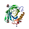 8f0vC  4nyqS S: Starting model for refinement C: citing same article ( |
|---|---|
| Similar structure data |
-Links
 PDBj
PDBj

- Assembly
| Deposited unit | 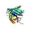
| ||||||||
|---|---|---|---|---|---|---|---|---|---|
| 1 |
| ||||||||
| Unit cell |
|
-Components
| #1: Protein | Mass: 17912.180 Da / Num. of mol.: 1 Source method: isolated from a genetically manipulated source Source: (gene. exp.) Diploptera punctata (Pacific beetle cockroach)Production host:  | ||||||
|---|---|---|---|---|---|---|---|
| #2: Sugar |   Type: D-saccharide, beta linking / Mass: 221.208 Da / Num. of mol.: 3 / Source method: obtained synthetically / Formula: C8H15NO6 Type: D-saccharide, beta linking / Mass: 221.208 Da / Num. of mol.: 3 / Source method: obtained synthetically / Formula: C8H15NO6#3: Water | ChemComp-HOH / |  Mass: 18.015 Da / Num. of mol.: 7 / Source method: isolated from a natural source / Formula: H2O Mass: 18.015 Da / Num. of mol.: 7 / Source method: isolated from a natural source / Formula: H2OHas ligand of interest | N | Has protein modification | Y | |
-Experimental details
-Experiment
| Experiment | Method: X-RAY DIFFRACTION / Number of used crystals: 1 |
|---|
- Sample preparation
| Crystal | Density Matthews: 2.19 Å3/Da / Density % sol: 43.74 % / Description: Rod shaped crystals |
|---|---|
| Crystal grow | Temperature: 293 K / Method: vapor diffusion, hanging drop / pH: 6.9 / Details: 0.2M Potassium Nitrate, pH 6.9, 20% PEG 3350 |
-Data collection
| Diffraction | Mean temperature: 100 K / Serial crystal experiment: N | |||||||||||||||||||||
|---|---|---|---|---|---|---|---|---|---|---|---|---|---|---|---|---|---|---|---|---|---|---|
| Diffraction source | Source: SYNCHROTRON / Site: APS  / Beamline: 23-ID-D / Wavelength: 1.0332 Å / Beamline: 23-ID-D / Wavelength: 1.0332 Å | |||||||||||||||||||||
| Detector | Type: DECTRIS PILATUS 6M / Detector: PIXEL / Date: Jul 21, 2022 | |||||||||||||||||||||
| Radiation | Monochromator: Double Crystal / Protocol: SINGLE WAVELENGTH / Monochromatic (M) / Laue (L): M / Scattering type: x-ray | |||||||||||||||||||||
| Radiation wavelength | Wavelength: 1.0332 Å / Relative weight: 1 | |||||||||||||||||||||
| Reflection | Resolution: 2.1→37.02 Å / Num. obs: 8498 / % possible obs: 96.7 % / Redundancy: 2.6 % / CC1/2: 0.98 / Rmerge(I) obs: 0.112 / Rpim(I) all: 0.112 / Rrim(I) all: 0.158 / Net I/σ(I): 3.5 | |||||||||||||||||||||
| Reflection shell | Diffraction-ID: 1 / Redundancy: 2.6 %
|
- Processing
| Software |
| |||||||||||||||||||||||||||||||||||||||||||||||||||||||||||||||||||||||||||||||||||||||||||||||||||||||||||||||||||||||||||||||||||||||||||||||||||||||||||||||||||||||||||||||||||||||||||||||||||||||||||||||||||||||||||||||||||||||
|---|---|---|---|---|---|---|---|---|---|---|---|---|---|---|---|---|---|---|---|---|---|---|---|---|---|---|---|---|---|---|---|---|---|---|---|---|---|---|---|---|---|---|---|---|---|---|---|---|---|---|---|---|---|---|---|---|---|---|---|---|---|---|---|---|---|---|---|---|---|---|---|---|---|---|---|---|---|---|---|---|---|---|---|---|---|---|---|---|---|---|---|---|---|---|---|---|---|---|---|---|---|---|---|---|---|---|---|---|---|---|---|---|---|---|---|---|---|---|---|---|---|---|---|---|---|---|---|---|---|---|---|---|---|---|---|---|---|---|---|---|---|---|---|---|---|---|---|---|---|---|---|---|---|---|---|---|---|---|---|---|---|---|---|---|---|---|---|---|---|---|---|---|---|---|---|---|---|---|---|---|---|---|---|---|---|---|---|---|---|---|---|---|---|---|---|---|---|---|---|---|---|---|---|---|---|---|---|---|---|---|---|---|---|---|---|---|---|---|---|---|---|---|---|---|---|---|---|---|---|---|---|---|
| Refinement | Method to determine structure: MOLECULAR REPLACEMENT Starting model: 4NYQ Resolution: 2.1→37.012 Å / Cor.coef. Fo:Fc: 0.943 / Cor.coef. Fo:Fc free: 0.921 / SU B: 9.06 / SU ML: 0.225 / Cross valid method: FREE R-VALUE / ESU R: 0.277 / ESU R Free: 0.216 Details: Hydrogens have been added in their riding positions
| |||||||||||||||||||||||||||||||||||||||||||||||||||||||||||||||||||||||||||||||||||||||||||||||||||||||||||||||||||||||||||||||||||||||||||||||||||||||||||||||||||||||||||||||||||||||||||||||||||||||||||||||||||||||||||||||||||||||
| Solvent computation | Ion probe radii: 0.8 Å / Shrinkage radii: 0.8 Å / VDW probe radii: 1.2 Å / Solvent model: MASK BULK SOLVENT | |||||||||||||||||||||||||||||||||||||||||||||||||||||||||||||||||||||||||||||||||||||||||||||||||||||||||||||||||||||||||||||||||||||||||||||||||||||||||||||||||||||||||||||||||||||||||||||||||||||||||||||||||||||||||||||||||||||||
| Displacement parameters | Biso mean: 40.13 Å2
| |||||||||||||||||||||||||||||||||||||||||||||||||||||||||||||||||||||||||||||||||||||||||||||||||||||||||||||||||||||||||||||||||||||||||||||||||||||||||||||||||||||||||||||||||||||||||||||||||||||||||||||||||||||||||||||||||||||||
| Refinement step | Cycle: LAST / Resolution: 2.1→37.012 Å
| |||||||||||||||||||||||||||||||||||||||||||||||||||||||||||||||||||||||||||||||||||||||||||||||||||||||||||||||||||||||||||||||||||||||||||||||||||||||||||||||||||||||||||||||||||||||||||||||||||||||||||||||||||||||||||||||||||||||
| Refine LS restraints |
| |||||||||||||||||||||||||||||||||||||||||||||||||||||||||||||||||||||||||||||||||||||||||||||||||||||||||||||||||||||||||||||||||||||||||||||||||||||||||||||||||||||||||||||||||||||||||||||||||||||||||||||||||||||||||||||||||||||||
| LS refinement shell | Refine-ID: X-RAY DIFFRACTION / Total num. of bins used: 20
|
