 Movie
Movie Controller

[English] 日本語
 Yorodumi
- PDB-8evk: Crystal structure of Helicobacter pylori dihydroneopterin aldolas... -
+ Open data

- Basic information
| Entry | Database: PDB / ID: 8evk | ||||||
|---|---|---|---|---|---|---|---|
| Title | Crystal structure of Helicobacter pylori dihydroneopterin aldolase (DHNA) | ||||||
 Components Components | Dihydroneopterin aldolase | ||||||
 Keywords Keywords | LYASE / Inhibitor / Complex / Aldolase | ||||||
| Function / homology |  Function and homology information Function and homology informationdihydroneopterin aldolase activity / folic acid-containing compound metabolic process Similarity search - Function | ||||||
| Biological species |  Helicobacter pylori G27 (bacteria) Helicobacter pylori G27 (bacteria) | ||||||
| Method |  X-RAY DIFFRACTION / SYNCHROTRON / MOLECULAR REPLACEMENT / Resolution: 1.49 Å X-RAY DIFFRACTION / SYNCHROTRON / MOLECULAR REPLACEMENT / Resolution: 1.49 Å | ||||||
 Authors Authors | Shaw, G.X. / Cherry, S. / Tropea, J.E. / Ji, X. | ||||||
| Funding support |  United States, 1items United States, 1items
| ||||||
 Citation Citation | Journal: Curr Res Struct Biol / Year: 2023 Title: Structure of Helicobacter pylori dihydroneopterin aldolase suggests a fragment-based strategy for isozyme-specific inhibitor design. Authors: Shaw, G.X. / Fan, L. / Cherry, S. / Shi, G. / Tropea, J.E. / Ji, X. | ||||||
| History |
|
- Structure visualization
| Structure viewer | Molecule: MolmilJmol/JSmol |
|---|
- Downloads & links
-Download
| PDBx/mmCIF format | 8evk.cif.gz | 45.4 KB | Display | PDBx/mmCIF format |
|---|---|---|---|---|
| PDB format | pdb8evk.ent.gz | 29.4 KB | Display | PDB format |
| PDBx/mmJSON format | 8evk.json.gz | Tree view | PDBx/mmJSON format | |
| Others |  Other downloads Other downloads |
-Validation report
| Arichive directory | https://data.pdbj.org/pub/pdb/validation_reports/ev/8evkftp://data.pdbj.org/pub/pdb/validation_reports/ev/8evk | HTTPS FTP |
|---|
-Related structure data
| Related structure data |  2o90S S: Starting model for refinement |
|---|---|
| Similar structure data |
-Links
 PDBj
PDBj
- Assembly
| Deposited unit | 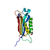
| ||||||||||||
|---|---|---|---|---|---|---|---|---|---|---|---|---|---|
| 1 | 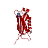
| ||||||||||||
| Unit cell |
| ||||||||||||
| Components on special symmetry positions |
|
-Components
| #1: Protein | Mass: 13953.307 Da / Num. of mol.: 1 / Fragment: Full-length Source method: isolated from a genetically manipulated source Source: (gene. exp.) Helicobacter pylori G27 (bacteria) / Strain: G27 / Gene: HPG27_1434 / Plasmid: pJT250 / Production host: |
|---|---|
| #2: Chemical | ChemComp-PE0 /   Mass: 163.137 Da / Num. of mol.: 1 / Source method: obtained synthetically / Formula: C6H5N5O / Feature type: SUBJECT OF INVESTIGATION Mass: 163.137 Da / Num. of mol.: 1 / Source method: obtained synthetically / Formula: C6H5N5O / Feature type: SUBJECT OF INVESTIGATION |
| #3: Chemical | ChemComp-EDO /   Mass: 62.068 Da / Num. of mol.: 1 / Source method: obtained synthetically / Formula: C2H6O2 Mass: 62.068 Da / Num. of mol.: 1 / Source method: obtained synthetically / Formula: C2H6O2 |
| #4: Water | ChemComp-HOH /  Mass: 18.015 Da / Num. of mol.: 134 / Source method: isolated from a natural source / Formula: H2O Mass: 18.015 Da / Num. of mol.: 134 / Source method: isolated from a natural source / Formula: H2O |
| Has ligand of interest | Y |
-Experimental details
-Experiment
| Experiment | Method: X-RAY DIFFRACTION / Number of used crystals: 1 |
|---|
- Sample preparation
| Crystal | Density Matthews: 2.42 Å3/Da / Density % sol: 49.24 % / Mosaicity: 1.601 ° / Mosaicity esd: 0.012 ° |
|---|---|
| Crystal grow | Temperature: 293 K / Method: vapor diffusion, sitting drop / pH: 7.5 / Details: (NH4)2SO4, NaCl, etc. |
-Data collection
| Diffraction | Mean temperature: 100 K / Serial crystal experiment: N | |||||||||||||||||||||||||||||||||||||||||||||||||||||||||||||||||||||||||||||||||||||||||||||||||||
|---|---|---|---|---|---|---|---|---|---|---|---|---|---|---|---|---|---|---|---|---|---|---|---|---|---|---|---|---|---|---|---|---|---|---|---|---|---|---|---|---|---|---|---|---|---|---|---|---|---|---|---|---|---|---|---|---|---|---|---|---|---|---|---|---|---|---|---|---|---|---|---|---|---|---|---|---|---|---|---|---|---|---|---|---|---|---|---|---|---|---|---|---|---|---|---|---|---|---|---|---|
| Diffraction source | Source: SYNCHROTRON / Site: APS / Beamline: 22-BM / Wavelength: 1 Å | |||||||||||||||||||||||||||||||||||||||||||||||||||||||||||||||||||||||||||||||||||||||||||||||||||
| Detector | Type: MARMOSAIC 225 mm CCD / Detector: CCD / Date: Mar 15, 2013 / Details: mirrors | |||||||||||||||||||||||||||||||||||||||||||||||||||||||||||||||||||||||||||||||||||||||||||||||||||
| Radiation | Monochromator: Double crystal / Protocol: SINGLE WAVELENGTH / Monochromatic (M) / Laue (L): M / Scattering type: x-ray | |||||||||||||||||||||||||||||||||||||||||||||||||||||||||||||||||||||||||||||||||||||||||||||||||||
| Radiation wavelength | Wavelength: 1 Å / Relative weight: 1 | |||||||||||||||||||||||||||||||||||||||||||||||||||||||||||||||||||||||||||||||||||||||||||||||||||
| Reflection | Resolution: 1.49→30 Å / Num. obs: 21197 / % possible obs: 98.2 % / Redundancy: 14.1 % / Biso Wilson estimate: 22.81 Å2 / CC1/2: 0.999 / CC star: 1 / Rmerge(I) obs: 0.132 / Rpim(I) all: 0.036 / Rrim(I) all: 0.136 / Χ2: 0.96 / Net I/av σ(I): 17.45 / Net I/σ(I): 13.7 | |||||||||||||||||||||||||||||||||||||||||||||||||||||||||||||||||||||||||||||||||||||||||||||||||||
| Reflection shell | Diffraction-ID: 1
|
- Processing
| Software |
| ||||||||||||||||||||||||||||||||||||||||||||||||||||||||
|---|---|---|---|---|---|---|---|---|---|---|---|---|---|---|---|---|---|---|---|---|---|---|---|---|---|---|---|---|---|---|---|---|---|---|---|---|---|---|---|---|---|---|---|---|---|---|---|---|---|---|---|---|---|---|---|---|---|
| Refinement | Method to determine structure: MOLECULAR REPLACEMENT Starting model: 2O90 Resolution: 1.49→26.9 Å / SU ML: 0.2427 / Cross valid method: FREE R-VALUE / σ(F): 1.37 / Phase error: 31.421 Stereochemistry target values: GeoStd + Monomer Library + CDL v1.2
| ||||||||||||||||||||||||||||||||||||||||||||||||||||||||
| Solvent computation | Shrinkage radii: 0.9 Å / VDW probe radii: 1.11 Å / Solvent model: FLAT BULK SOLVENT MODEL | ||||||||||||||||||||||||||||||||||||||||||||||||||||||||
| Displacement parameters | Biso mean: 30.63 Å2 | ||||||||||||||||||||||||||||||||||||||||||||||||||||||||
| Refinement step | Cycle: LAST / Resolution: 1.49→26.9 Å
| ||||||||||||||||||||||||||||||||||||||||||||||||||||||||
| Refine LS restraints |
| ||||||||||||||||||||||||||||||||||||||||||||||||||||||||
| LS refinement shell |
|
