National Institutes of Health/National Institute of General Medical Sciences (NIH/NIGMS)
P41GM136508
United States
Howard Hughes Medical Institute (HHMI)
United States
Citation
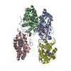
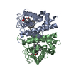
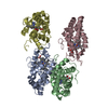
Journal: J Am Chem Soc / Year: 2023 Title: MicroED Structure of a Protoglobin Reactive Carbene Intermediate. Authors: Emma Danelius / Nicholas J Porter / Johan Unge / Frances H Arnold / Tamir Gonen / Abstract: Microcrystal electron diffraction (MicroED) is an emerging technique that has shown great potential for describing new chemical and biological molecular structures. Several important structures of ...Microcrystal electron diffraction (MicroED) is an emerging technique that has shown great potential for describing new chemical and biological molecular structures. Several important structures of small molecules, natural products, and peptides have been determined using methods. However, only a couple of novel protein structures have thus far been derived by MicroED. Taking advantage of recent technological advances, including higher acceleration voltage and using a low-noise detector in counting mode, we have determined the first structure of an protoglobin (Pgb) variant by MicroED using an AlphaFold2 model for phasing. The structure revealed that mutations introduced during directed evolution enhance carbene transfer activity by reorienting an α helix of Pgb into a dynamic loop, making the catalytic active site more readily accessible. After exposing the tiny crystals to the substrate, we also trapped the reactive iron-carbenoid intermediate involved in this engineered Pgb's new-to-nature activity, a challenging carbene transfer from a diazirine via a putative metallo-carbene. The bound structure discloses how an enlarged active site pocket stabilizes the carbene bound to the heme iron and, presumably, the transition state for the formation of this key intermediate. This work demonstrates that improved MicroED technology and the advancement in protein structure prediction now enable investigation of structures that was previously beyond reach.
 Movie
Movie Controller


 Open data
 Basic information
 Components
 Keywords
 Function and homology information
 Aeropyrum pernix (archaea)
 Authors
 United States, 2items
  Citation
 Structure visualization
 Downloads & links
 Other downloads

 PDBj
PDBj


 Assembly