[English] 日本語
Yorodumi
- PDB-7ute: MicroED structure of Aeropyrum pernix protoglobin mutant -

+
Open data


ID or keywords:

Loading...

-
Basic information

Entry
Database: PDB / ID: 7ute
TitleMicroED structure of Aeropyrum pernix protoglobin mutant
ComponentsProtogloblin ApPgb
KeywordsMETAL BINDING PROTEIN / MicroED / protoglobin / heme
Function / homology
Function and homology information


oxygen binding / heme binding
Similarity search - Function
Protoglobin / Globin-sensor domain / Protoglobin / Globin/Protoglobin / Globin-like superfamily
Similarity search - Domain/homology
PROTOPORPHYRIN IX CONTAINING FE / IMIDAZOLE / Protogloblin ApPgb
Similarity search - Component
Biological speciesAeropyrum pernix (archaea)
MethodELECTRON CRYSTALLOGRAPHY / electron crystallography / MOLECULAR REPLACEMENT / cryo EM / Resolution: 2.1 Å
AuthorsDanelius, E. / Gonen, T. / Unge, J.T.
Funding support United States, 2items
OrganizationGrant numberCountry
National Institutes of Health/National Institute of General Medical Sciences (NIH/NIGMS)P41GM136508 United States
Howard Hughes Medical Institute (HHMI) United States
Citation
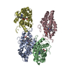
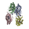
Journal: J Am Chem Soc / Year: 2022
Title: Biocatalytic Carbene Transfer Using Diazirines.
Authors: Nicholas J Porter / Emma Danelius / Tamir Gonen / Frances H Arnold /
Abstract: Biocatalytic carbene transfer from diazo compounds is a versatile strategy in asymmetric synthesis. However, the limited pool of stable diazo compounds constrains the variety of accessible products. ...Biocatalytic carbene transfer from diazo compounds is a versatile strategy in asymmetric synthesis. However, the limited pool of stable diazo compounds constrains the variety of accessible products. To overcome this restriction, we have engineered variants of protoglobin (Pgb) that use diazirines as carbene precursors. While the enhanced stability of diazirines relative to their diazo isomers enables access to a diverse array of carbenes, they have previously resisted catalytic activation. Our engineered Pgb variants represent the first example of catalysts for selective carbene transfer from these species at room temperature. The structure of an Pgb variant, determined by microcrystal electron diffraction (MicroED), reveals that evolution has enhanced access to the heme active site to facilitate this new-to-nature catalysis. Using readily prepared aryl diazirines as model substrates, we demonstrate the application of these highly stable carbene precursors in biocatalytic cyclopropanation, N-H insertion, and Si-H insertion reactions.
History
DepositionApr 26, 2022Deposition site: RCSB / Processing site: RCSB
Revision 1.0May 25, 2022Provider: repository / Type: Initial release
Revision 1.1Jun 8, 2022Group: Database references / Category: citation / citation_author
Item: _citation.journal_volume / _citation.page_first ..._citation.journal_volume / _citation.page_first / _citation.page_last / _citation_author.identifier_ORCID
Revision 1.2Feb 14, 2024Group: Data collection / Category: chem_comp_atom / chem_comp_bond

-
Structure visualization

Structure viewerMolecule:
MolmilJmol/JSmol

Downloads & links

-
Assembly

Deposited unit
A: Protogloblin ApPgb
B: Protogloblin ApPgb
C: Protogloblin ApPgb
D: Protogloblin ApPgb
hetero molecules


Theoretical massNumber of molelcules
Total (without water)93,61812
Polymers90,8764
Non-polymers2,7428
Water4,324240
1


  • Idetical with deposited unit
  • defined by author
TypeNameSymmetry operationNumber
identity operation1_555x,y,z1
Unit cell
Length a, b, c (Å)46.246, 58.305, 80.732
Angle α, β, γ (deg.)104.080, 98.590, 90.100
Int Tables number1
Space group name H-MP1
Space group name HallP1
Symmetry operation#1: x,y,z
Noncrystallographic symmetry (NCS)NCS domain:
IDEns-IDDetails
d_1ens_1(chain "D" and (resid 6 through 12 or resid 14...
d_2ens_1(chain "B" and (resid 6 through 12 or resid 14...
d_3ens_1(chain "C" and (resid 6 through 12 or resid 14...
d_4ens_1(chain "A" and (resid 6 through 12 or resid 14...

NCS domain segments:
Dom-IDComponent-IDEns-IDBeg label comp-IDEnd label comp-IDLabel asym-IDLabel seq-ID
d_11ens_1ILEGLYD1 - 7
d_12ens_1VALLYSD9 - 23
d_13ens_1THRGLYD25 - 29
d_14ens_1LYSLEUD31 - 43
d_15ens_1ASPASPD45 - 48
d_16ens_1ILEGLYD50 - 56
d_17ens_1ILELYSD59 - 72
d_18ens_1TYRLEUD74 - 75
d_19ens_1ARGSERD77 - 112
d_110ens_1LYSTRPD114 - 184
d_111ens_1IMDIMDD
d_21ens_1ILEGLYB1 - 7
d_22ens_1VALLYSB9 - 23
d_23ens_1THRGLYB25 - 29
d_24ens_1LYSLEUB31 - 43
d_25ens_1ASPASPB45 - 48
d_26ens_1ILEGLYB50 - 56
d_27ens_1ILELYSB59 - 72
d_28ens_1TYRLEUB74 - 75
d_29ens_1ARGSERB77 - 112
d_210ens_1LYSTRPB114 - 184
d_211ens_1IMDIMDB
d_31ens_1ILEGLYC1 - 7
d_32ens_1VALLYSC9 - 23
d_33ens_1THRGLYC25 - 29
d_34ens_1LYSLEUC31 - 43
d_35ens_1ASPASPC45 - 48
d_36ens_1ILEGLYC50 - 56
d_37ens_1ILELYSC59 - 72
d_38ens_1TYRLEUC74 - 75
d_39ens_1ARGSERC77 - 112
d_310ens_1LYSTRPC114 - 184
d_311ens_1IMDIMDC
d_41ens_1ILEGLYA1 - 7
d_42ens_1VALLYSA9 - 23
d_43ens_1THRGLYA25 - 29
d_44ens_1LYSLEUA31 - 43
d_45ens_1ASPASPA45 - 48
d_46ens_1ILEGLYA50 - 56
d_47ens_1ILELYSA65 - 78
d_48ens_1TYRLEUA80 - 81
d_49ens_1ARGSERA83 - 118
d_410ens_1LYSTRPA120 - 190
d_411ens_1IMDIMDA

NCS oper:
IDCodeMatrixVector
1given(-0.999994587624, -0.00317152480588, 0.00087530122274), (-0.0031792974112, 0.999954209038, -0.00902617835407), (-0.000846634393303, -0.00902891234392, -0.999958880131)11.5922099593, -29.2646444914, -34.0527349825
2given(-0.9999984669, -0.00165338130169, 0.000576653175149), (0.00165651335553, -0.999983645944, 0.00547392065957), (0.000567593266465, 0.00547486750119, 0.999984851717)34.7006855008, -17.3082588887, -0.0551359046483
3given(0.999998869961, 0.00146414541039, -0.000341109745264), (0.00146476231479, -0.999997279491, 0.0018153464962), (-0.000338450886031, -0.00181584408949, -0.999998294079)-23.1326103095, 11.801868876, -34.1907522948

-
Components

#1: Protein
Protogloblin ApPgb


Mass: 22718.887 Da / Num. of mol.: 4
Source method: isolated from a genetically manipulated source
Source: (gene. exp.) Aeropyrum pernix (archaea) / Gene: APE_0287 / Production host: Escherichia coli (E. coli) / References: UniProt: Q9YFF4
#2: Chemical
ChemComp-HEM / PROTOPORPHYRIN IX CONTAINING FE / HEME / Heme B


Mass: 616.487 Da / Num. of mol.: 4 / Source method: obtained synthetically / Formula: C34H32FeN4O4 / Feature type: SUBJECT OF INVESTIGATION
#3: Chemical
ChemComp-IMD / IMIDAZOLE / Imidazole


Mass: 69.085 Da / Num. of mol.: 4 / Source method: obtained synthetically / Formula: C3H5N2
#4: Water ChemComp-HOH / water / Water


Mass: 18.015 Da / Num. of mol.: 240 / Source method: isolated from a natural source / Formula: H2O
Has ligand of interestY

-
Experimental details

-
Experiment

ExperimentMethod: ELECTRON CRYSTALLOGRAPHY
EM experimentAggregation state: 3D ARRAY / 3D reconstruction method: electron crystallography

-
Sample preparation

ComponentName: Homotetramer of Aeropyrum pernix protoglobin (ApePgb) C45G, W59L, Y60V, V63R, C102S, F145Q, I149L mutant
Type: COMPLEX / Entity ID: #1 / Source: RECOMBINANT
Source (natural)Organism: Aeropyrum pernix (archaea)
Source (recombinant)Organism: Escherichia coli (E. coli)
Buffer solutionpH: 8
SpecimenConc.: 20 mg/ml / Embedding applied: NO / Shadowing applied: NO / Staining applied: NO / Vitrification applied: YES
Specimen supportGrid material: COPPER / Grid type: Quantifoil R2/2
VitrificationInstrument: LEICA EM GP / Cryogen name: ETHANE / Humidity: 90 % / Chamber temperature: 277 K

-
Data collection

Experimental equipment
Model: Titan Krios / Image courtesy: FEI Company
MicroscopyModel: FEI TITAN KRIOS
Electron gunElectron source: FIELD EMISSION GUN / Accelerating voltage: 300 kV / Illumination mode: OTHER
Electron lensMode: DIFFRACTION / Nominal defocus max: 0 nm / Nominal defocus min: 0 nm / Cs: 2.7 mm / C2 aperture diameter: 50 µm / Alignment procedure: BASIC
Specimen holderCryogen: NITROGEN / Specimen holder model: FEI TITAN KRIOS AUTOGRID HOLDER / Temperature (max): 90 K / Temperature (min): 77 K
Image recordingElectron dose: 0.00025 e/Å2 / Detector mode: COUNTING / Film or detector model: FEI FALCON IV (4k x 4k)
Image scansSampling size: 14 µm / Width: 4096 / Height: 4096
EM diffractionCamera length: 2460 mm
EM diffraction shellResolution: 2.1→24.26 Å / Fourier space coverage: 72 % / Multiplicity: 4.1 / Num. of structure factors: 33825 / Phase residual: 29.25 °
EM diffraction statsFourier space coverage: 72 % / High resolution: 2.1 Å / Num. of intensities measured: 33825 / Num. of structure factors: 33825 / Phase error rejection criteria: not applicable / Rmerge: 0.43
DiffractionSerial crystal experiment: N
DetectorDate: Mar 29, 2022
ReflectionResolution: 2.1→23.391 Å / Num. obs: 34624 / Biso Wilson estimate: 8.58 Å2 / CC1/2: 0.972
Reflection shellResolution: 2.1→2.15 Å / Mean I/σ(I) obs: 2.4 / Num. unique obs: 2481 / CC1/2: 0.768 / % possible all: 71

-
Processing

Software
NameVersionClassificationNB
PHENIX1.20.1_4487refinement
XDSdata reduction
Aimlessdata scaling
PHASERphasing
EM softwareName: PHENIX / Version: 1.20.1-4487-000 / Category: 3D reconstruction
EM 3D crystal entity∠α: 104.1 ° / ∠β: 98.6 ° / ∠γ: 90.1 ° / A: 46.2 Å / B: 58.3 Å / C: 80.7 Å / Space group name: P1 / Space group num: 1
CTF correctionType: NONE
3D reconstructionResolution method: DIFFRACTION PATTERN/LAYERLINES / Symmetry type: 3D CRYSTAL
Atomic model buildingB value: 18.2 / Protocol: OTHER / Space: RECIPROCAL / Target criteria: maximum likelihood
RefinementMethod to determine structure: MOLECULAR REPLACEMENT
Starting model: predicted model from AlphaFold

Resolution: 2.1→24.26 Å / SU ML: 0.2339 / Cross valid method: FREE R-VALUE / σ(F): 1.96 / Phase error: 20.8958
Stereochemistry target values: GeoStd + Monomer Library + CDL v1.2
RfactorNum. reflection% reflection
Rfree0.2209 1632 4.82 %
Rwork0.1972 32193 -
obs0.1983 33825 71.77 %
Solvent computationShrinkage radii: 0.9 Å / VDW probe radii: 1.1 Å / Solvent model: FLAT BULK SOLVENT MODEL
Displacement parametersBiso mean: 18.16 Å2
Refinement stepCycle: LAST / Resolution: 2.1→24.26 Å
ProteinNucleic acidLigandSolventTotal
Num. atoms6108 0 192 240 6540
Refine LS restraints
Refine-IDTypeDev idealNumber
ELECTRON CRYSTALLOGRAPHYf_bond_d0.01326504
ELECTRON CRYSTALLOGRAPHYf_angle_d1.55798881
ELECTRON CRYSTALLOGRAPHYf_chiral_restr0.0679910
ELECTRON CRYSTALLOGRAPHYf_plane_restr0.0121104
ELECTRON CRYSTALLOGRAPHYf_dihedral_angle_d17.1241865
Refine LS restraints NCS
Ens-IDDom-IDAuth asym-IDRefine-IDTypeRms dev position (Å)
ens_1d_2BELECTRON CRYSTALLOGRAPHYNCS constraints0.0288166087992
ens_1d_3CELECTRON CRYSTALLOGRAPHYNCS constraints0.0394672407305
ens_1d_4AELECTRON CRYSTALLOGRAPHYNCS constraints0.0327778333716
LS refinement shell
Resolution (Å)Rfactor RfreeNum. reflection RfreeRfactor RworkNum. reflection RworkRefine-ID% reflection obs (%)
2.1-2.160.2471370.23172500ELECTRON CRYSTALLOGRAPHY67.79
2.16-2.230.27921360.2322613ELECTRON CRYSTALLOGRAPHY69.23
2.23-2.310.26181350.23242630ELECTRON CRYSTALLOGRAPHY70.27
2.31-2.40.28421400.22372596ELECTRON CRYSTALLOGRAPHY70.24
2.4-2.510.26741320.23142661ELECTRON CRYSTALLOGRAPHY71.16
2.51-2.650.23511410.22092725ELECTRON CRYSTALLOGRAPHY72.69
2.65-2.810.21941160.20872738ELECTRON CRYSTALLOGRAPHY72.88
2.81-3.030.25591370.20712772ELECTRON CRYSTALLOGRAPHY74.04
3.03-3.330.21111540.18942773ELECTRON CRYSTALLOGRAPHY74.48
3.33-3.810.18231320.16362795ELECTRON CRYSTALLOGRAPHY74.52
3.81-4.80.14321350.14182739ELECTRON CRYSTALLOGRAPHY73.22
Refinement TLS params.Method: refined / Origin x: 5.85169117988 Å / Origin y: -8.64639316368 Å / Origin z: -16.889422311 Å
111213212223313233
T0.0162048581633 Å20.00579909235784 Å2-0.0227890380203 Å2-0.027729296648 Å20.00386312842991 Å2--0.0167965738518 Å2
L0.291819134287 °20.0548320643967 °2-0.0604016817159 °2-0.337960531343 °20.0100386789426 °2--0.0575517009793 °2
S0.0149261756223 Å °-0.00837558580237 Å °-0.00744721659688 Å °0.0176035253845 Å °-0.0176661579133 Å °-0.00231255884188 Å °0.00291278070697 Å °0.000768863252757 Å °-0.0153236069714 Å °
Refinement TLS groupSelection details: all

+
About Yorodumi

-
News

-
Feb 9, 2022. New format data for meta-information of EMDB entries

New format data for meta-information of EMDB entries

  • Version 3 of the EMDB header file is now the official format.
  • The previous official version 1.9 will be removed from the archive.

Related info.:EMDB header

External links:wwPDB to switch to version 3 of the EMDB data model

-
Aug 12, 2020. Covid-19 info

Covid-19 info

URL: https://pdbj.org/emnavi/covid19.php

New page: Covid-19 featured information page in EM Navigator.

Related info.:Covid-19 info / Mar 5, 2020. Novel coronavirus structure data

+
Mar 5, 2020. Novel coronavirus structure data

Novel coronavirus structure data

Related info.:Yorodumi Speices / Aug 12, 2020. Covid-19 info

External links:COVID-19 featured content - PDBj / Molecule of the Month (242):Coronavirus Proteases

+
Jan 31, 2019. EMDB accession codes are about to change! (news from PDBe EMDB page)

EMDB accession codes are about to change! (news from PDBe EMDB page)

  • The allocation of 4 digits for EMDB accession codes will soon come to an end. Whilst these codes will remain in use, new EMDB accession codes will include an additional digit and will expand incrementally as the available range of codes is exhausted. The current 4-digit format prefixed with “EMD-” (i.e. EMD-XXXX) will advance to a 5-digit format (i.e. EMD-XXXXX), and so on. It is currently estimated that the 4-digit codes will be depleted around Spring 2019, at which point the 5-digit format will come into force.
  • The EM Navigator/Yorodumi systems omit the EMD- prefix.

Related info.:Q: What is EMD? / ID/Accession-code notation in Yorodumi/EM Navigator

External links:EMDB Accession Codes are Changing Soon! / Contact to PDBj

+
Jul 12, 2017. Major update of PDB

Major update of PDB

  • wwPDB released updated PDB data conforming to the new PDBx/mmCIF dictionary.
  • This is a major update changing the version number from 4 to 5, and with Remediation, in which all the entries are updated.
  • In this update, many items about electron microscopy experimental information are reorganized (e.g. em_software).
  • Now, EM Navigator and Yorodumi are based on the updated data.

External links:wwPDB Remediation / Enriched Model Files Conforming to OneDep Data Standards Now Available in the PDB FTP Archive

-
Yorodumi

Thousand views of thousand structures

  • Yorodumi is a browser for structure data from EMDB, PDB, SASBDB, etc.
  • This page is also the successor to EM Navigator detail page, and also detail information page/front-end page for Omokage search.
  • The word "yorodu" (or yorozu) is an old Japanese word meaning "ten thousand". "mi" (miru) is to see.

Related info.:EMDB / PDB / SASBDB / Comparison of 3 databanks / Yorodumi Search / Aug 31, 2016. New EM Navigator & Yorodumi / Yorodumi Papers / Jmol/JSmol / Function and homology information / Changes in new EM Navigator and Yorodumi

Read more