 Movie
Movie Controller


+ Open data

- Basic information
| Entry | Database: PDB / ID: 8esv | |||||||||
|---|---|---|---|---|---|---|---|---|---|---|
| Title | Structure of human ADAM10-Tspan15 complex bound to 11G2 vFab | |||||||||
 Components Components |
| |||||||||
 Keywords Keywords | MEMBRANE PROTEIN / Protease / Metalloprotease / Tetraspanin / Sheddase / Adhesion | |||||||||
| Function / homology |  Function and homology information Function and homology informationregulation of membrane protein ectodomain proteolysis / ADAM10 endopeptidase / constitutive protein ectodomain proteolysis / regulation of vasculature development / epidermal growth factor receptor ligand maturation / monocyte activation / metalloendopeptidase activity involved in amyloid precursor protein catabolic process / protein catabolic process at postsynapse / Constitutive Signaling by NOTCH1 t(7;9)(NOTCH1:M1580_K2555) Translocation Mutant / postsynapse organization ...regulation of membrane protein ectodomain proteolysis / ADAM10 endopeptidase / constitutive protein ectodomain proteolysis / regulation of vasculature development / epidermal growth factor receptor ligand maturation / monocyte activation / metalloendopeptidase activity involved in amyloid precursor protein catabolic process / protein catabolic process at postsynapse / Constitutive Signaling by NOTCH1 t(7;9)(NOTCH1:M1580_K2555) Translocation Mutant / postsynapse organization / perinuclear endoplasmic reticulum / tetraspanin-enriched microdomain / regulation of Notch signaling pathway / positive regulation of T cell chemotaxis / NOTCH4 Activation and Transmission of Signal to the Nucleus / pore complex assembly / metallodipeptidase activity / negative regulation of cell adhesion / adherens junction organization / positive regulation of tumor necrosis factor-mediated signaling pathway / clathrin-coated vesicle / regulation of postsynapse organization / Golgi-associated vesicle / amyloid precursor protein catabolic process / Signaling by EGFR / pore complex / regulation of neurotransmitter receptor localization to postsynaptic specialization membrane / Collagen degradation / membrane protein ectodomain proteolysis / tertiary granule membrane / negative regulation of Notch signaling pathway / extracellular matrix disassembly / cochlea development / response to tumor necrosis factor / EPH-ephrin mediated repulsion of cells / Notch signaling pathway / specific granule membrane / Degradation of the extracellular matrix / Constitutive Signaling by NOTCH1 HD Domain Mutants / NOTCH2 Activation and Transmission of Signal to the Nucleus / integrin-mediated signaling pathway / protein localization to plasma membrane / Activated NOTCH1 Transmits Signal to the Nucleus / synaptic membrane / Post-translational protein phosphorylation / adherens junction / NOTCH3 Activation and Transmission of Signal to the Nucleus / protein maturation / protein processing / metalloendopeptidase activity / SH3 domain binding / integrin binding / Regulation of Insulin-like Growth Factor (IGF) transport and uptake by Insulin-like Growth Factor Binding Proteins (IGFBPs) / Constitutive Signaling by NOTCH1 PEST Domain Mutants / Constitutive Signaling by NOTCH1 HD+PEST Domain Mutants / epidermal growth factor receptor signaling pathway / metallopeptidase activity / cell junction / positive regulation of tumor necrosis factor production / cell-cell signaling / late endosome membrane / in utero embryonic development / positive regulation of cell growth / endopeptidase activity / postsynaptic density / nuclear body / positive regulation of cell migration / endoplasmic reticulum lumen / Amyloid fiber formation / Golgi membrane / negative regulation of gene expression / axon / focal adhesion / positive regulation of cell population proliferation / Neutrophil degranulation / dendrite / glutamatergic synapse / Golgi apparatus / enzyme binding / cell surface / protein homodimerization activity / extracellular exosome / membrane / metal ion binding / plasma membrane / cytosol Similarity search - Function | |||||||||
| Biological species |  Homo sapiens (human) Homo sapiens (human) | |||||||||
| Method | ELECTRON MICROSCOPY / single particle reconstruction / cryo EM / Resolution: 3.3 Å | |||||||||
 Authors Authors | Lipper, C.H. / Blacklow, S.C. | |||||||||
| Funding support |  United States, 2items United States, 2items
| |||||||||
 Citation Citation | Journal: Cell / Year: 2023 Title: Structural basis for membrane-proximal proteolysis of substrates by ADAM10. Authors: Colin H Lipper / Emily D Egan / Khal-Hentz Gabriel / Stephen C Blacklow / Abstract: The endopeptidase ADAM10 is a critical catalyst for the regulated proteolysis of key drivers of mammalian development, physiology, and non-amyloidogenic cleavage of APP as the primary α-secretase. ...The endopeptidase ADAM10 is a critical catalyst for the regulated proteolysis of key drivers of mammalian development, physiology, and non-amyloidogenic cleavage of APP as the primary α-secretase. ADAM10 function requires the formation of a complex with a C8-tetraspanin protein, but how tetraspanin binding enables positioning of the enzyme active site for membrane-proximal cleavage remains unknown. We present here a cryo-EM structure of a vFab-ADAM10-Tspan15 complex, which shows that Tspan15 binding relieves ADAM10 autoinhibition and acts as a molecular measuring stick to position the enzyme active site about 20 Å from the plasma membrane for membrane-proximal substrate cleavage. Cell-based assays of N-cadherin shedding establish that the positioning of the active site by the interface between the ADAM10 catalytic domain and the bound tetraspanin influences selection of the preferred cleavage site. Together, these studies reveal the molecular mechanism underlying ADAM10 proteolysis at membrane-proximal sites and offer a roadmap for its modulation in disease. | |||||||||
| History |
|
- Structure visualization
| Structure viewer | Molecule: MolmilJmol/JSmol |
|---|
- Downloads & links
-Download
| PDBx/mmCIF format | 8esv.cif.gz | 207.2 KB | Display | PDBx/mmCIF format |
|---|---|---|---|---|
| PDB format | pdb8esv.ent.gz | 153.8 KB | Display | PDB format |
| PDBx/mmJSON format | 8esv.json.gz | Tree view | PDBx/mmJSON format | |
| Others |  Other downloads Other downloads |
-Validation report
| Arichive directory | https://data.pdbj.org/pub/pdb/validation_reports/es/8esvftp://data.pdbj.org/pub/pdb/validation_reports/es/8esv | HTTPS FTP |
|---|
-Related structure data
| Related structure data | 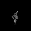 28580MC M: map data used to model this data C: citing same article ( |
|---|---|
| Similar structure data |
-Links
 PDBj
PDBj


- Assembly
| Deposited unit | 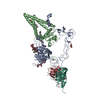
|
|---|---|
| 1 |
|
-Components
-Protein , 2 types, 2 molecules AB
| #1: Protein | Mass: 60477.340 Da / Num. of mol.: 1 Source method: isolated from a genetically manipulated source Source: (gene. exp.) Homo sapiens (human) / Gene: ADAM10, KUZ, MADM / Plasmid: pRK5M / Cell line (production host): Expi293F / Production host: Homo sapiens (human) / References: UniProt: O14672, ADAM10 endopeptidase |
|---|---|
| #2: Protein | Mass: 34740.469 Da / Num. of mol.: 1 Source method: isolated from a genetically manipulated source Source: (gene. exp.) Homo sapiens (human) / Gene: TSPAN15, NET7, TM4SF15, UNQ677/PRO1311 / Plasmid: pcDNA3.1/Hygro(+) / Cell line (production host): Expi293F / Production host: Homo sapiens (human) / References: UniProt: O95858 |
-Antibody , 2 types, 2 molecules HL
| #3: Antibody | Mass: 24135.105 Da / Num. of mol.: 1 Source method: isolated from a genetically manipulated source Source: (gene. exp.) Details (production host): Heavy and light chain in same vector with P2A sequence Cell line (production host): Expi293F / Production host: Homo sapiens (human) |
|---|---|
| #4: Antibody | Mass: 24185.803 Da / Num. of mol.: 1 Source method: isolated from a genetically manipulated source Source: (gene. exp.) Details (production host): Heavy and light chain in same vector with P2A sequence Cell line (production host): Expi293F / Production host: Homo sapiens (human) |
-Sugars , 3 types, 3 molecules 
| #5: Polysaccharide | alpha-D-mannopyranose-(1-3)-alpha-D-mannopyranose-(1-4)-2-acetamido-2-deoxy-beta-D-glucopyranose-(1- ...alpha-D-mannopyranose-(1-3)-alpha-D-mannopyranose-(1-4)-2-acetamido-2-deoxy-beta-D-glucopyranose-(1-4)-2-acetamido-2-deoxy-beta-D-glucopyranose Source method: isolated from a genetically manipulated source |
|---|---|
| #6: Polysaccharide | alpha-D-mannopyranose-(1-4)-2-acetamido-2-deoxy-beta-D-glucopyranose-(1-4)-2-acetamido-2-deoxy-beta- ...alpha-D-mannopyranose-(1-4)-2-acetamido-2-deoxy-beta-D-glucopyranose-(1-4)-2-acetamido-2-deoxy-beta-D-glucopyranose Source method: isolated from a genetically manipulated source |
| #7: Sugar | ChemComp-NAG /  Type: D-saccharide, beta linking / Mass: 221.208 Da / Num. of mol.: 1 / Source method: obtained synthetically / Formula: C8H15NO6 Type: D-saccharide, beta linking / Mass: 221.208 Da / Num. of mol.: 1 / Source method: obtained synthetically / Formula: C8H15NO6 |
-Non-polymers , 4 types, 5 molecules 


| #8: Chemical | ChemComp-CA /  Mass: 40.078 Da / Num. of mol.: 1 / Source method: obtained synthetically / Formula: Ca Mass: 40.078 Da / Num. of mol.: 1 / Source method: obtained synthetically / Formula: Ca |
|---|---|
| #9: Chemical | ChemComp-ZN /  Mass: 65.409 Da / Num. of mol.: 1 / Source method: obtained synthetically / Formula: Zn Mass: 65.409 Da / Num. of mol.: 1 / Source method: obtained synthetically / Formula: Zn |
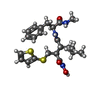
| #10: Chemical | ChemComp-BAT /  Mass: 477.640 Da / Num. of mol.: 1 / Source method: obtained synthetically / Formula: C23H31N3O4S2 / Comment: inhibitor*YM Mass: 477.640 Da / Num. of mol.: 1 / Source method: obtained synthetically / Formula: C23H31N3O4S2 / Comment: inhibitor*YM |
| #11: Chemical |  Mass: 486.726 Da / Num. of mol.: 2 / Source method: obtained synthetically / Formula: C31H50O4 Mass: 486.726 Da / Num. of mol.: 2 / Source method: obtained synthetically / Formula: C31H50O4 |
-Details
| Has ligand of interest | N |
|---|---|
| Has protein modification | Y |
-Experimental details
-Experiment
| Experiment | Method: ELECTRON MICROSCOPY |
|---|---|
| EM experiment | Aggregation state: PARTICLE / 3D reconstruction method: single particle reconstruction |
- Sample preparation
| Component |
| |||||||||||||||||||||||||
|---|---|---|---|---|---|---|---|---|---|---|---|---|---|---|---|---|---|---|---|---|---|---|---|---|---|---|
| Molecular weight | Experimental value: NO | |||||||||||||||||||||||||
| Source (natural) |
| |||||||||||||||||||||||||
| Source (recombinant) |
| |||||||||||||||||||||||||
| Buffer solution | pH: 7.4 | |||||||||||||||||||||||||
| Buffer component |
| |||||||||||||||||||||||||
| Specimen | Conc.: 2.1 mg/ml / Embedding applied: NO / Shadowing applied: NO / Staining applied: NO / Vitrification applied: YES / Details: This sample was monodisperse | |||||||||||||||||||||||||
| Specimen support | Grid material: COPPER / Grid mesh size: 400 divisions/in. / Grid type: Quantifoil | |||||||||||||||||||||||||
| Vitrification | Instrument: FEI VITROBOT MARK IV / Cryogen name: ETHANE / Humidity: 100 % / Chamber temperature: 295 K / Details: Blot for 7 seconds with a blot force of 15 |
- Electron microscopy imaging
| Experimental equipment |  Model: Titan Krios / Image courtesy: FEI Company |
|---|---|
| Microscopy | Model: FEI TITAN KRIOS |
| Electron gun | Electron source:  FIELD EMISSION GUN / Accelerating voltage: 300 kV / Illumination mode: FLOOD BEAM FIELD EMISSION GUN / Accelerating voltage: 300 kV / Illumination mode: FLOOD BEAM |
| Electron lens | Mode: BRIGHT FIELD / Nominal magnification: 105000 X / Nominal defocus max: 2200 nm / Nominal defocus min: 1000 nm / Cs: 2.7 mm / C2 aperture diameter: 50 µm |
| Specimen holder | Specimen holder model: FEI TITAN KRIOS AUTOGRID HOLDER |
| Image recording | Average exposure time: 1.3 sec. / Electron dose: 51.99 e/Å2 / Film or detector model: GATAN K3 BIOQUANTUM (6k x 4k) / Num. of grids imaged: 1 / Num. of real images: 10037 |
- Processing
| EM software |
| ||||||||||||||||||||||||||||||||||||||||
|---|---|---|---|---|---|---|---|---|---|---|---|---|---|---|---|---|---|---|---|---|---|---|---|---|---|---|---|---|---|---|---|---|---|---|---|---|---|---|---|---|---|
| CTF correction | Type: PHASE FLIPPING AND AMPLITUDE CORRECTION | ||||||||||||||||||||||||||||||||||||||||
| Symmetry | Point symmetry: C1 (asymmetric) | ||||||||||||||||||||||||||||||||||||||||
| 3D reconstruction | Resolution: 3.3 Å / Resolution method: FSC 0.143 CUT-OFF / Num. of particles: 178031 / Symmetry type: POINT | ||||||||||||||||||||||||||||||||||||||||
| Atomic model building | Space: REAL |
