 Movie
Movie Controller


+ Open data

- Basic information
| Entry | Database: PDB / ID: 8es4 | |||||||||||||||
|---|---|---|---|---|---|---|---|---|---|---|---|---|---|---|---|---|
| Title | Focused reconstruction of HRP29 tail | |||||||||||||||
 Components Components |
| |||||||||||||||
 Keywords Keywords | VIRUS / HRP29 | |||||||||||||||
| Function / homology |  Function and homology information Function and homology informationsymbiont genome ejection through host cell envelope, short tail mechanism / virus tail / outer membrane / virion component Similarity search - Function | |||||||||||||||
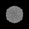
| Biological species |  Shigella phage Buco (virus) Shigella phage Buco (virus) | |||||||||||||||
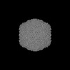
| Method | ELECTRON MICROSCOPY / single particle reconstruction / cryo EM / Resolution: 3.3 Å | |||||||||||||||
 Authors Authors | Subramanian, S. / Bergland Drarvik, S.M. / Parent, K.N. | |||||||||||||||
| Funding support |  United States, 4items United States, 4items
| |||||||||||||||
 Citation Citation | Journal: To Be Published Title: Structure of Shigella bacteriophage HRP29 Authors: Subramanian, S. / Bergland Drarvik, S.M. / Parent, K.N. | |||||||||||||||
| History |
|
- Structure visualization
| Structure viewer | Molecule: MolmilJmol/JSmol |
|---|
- Downloads & links
-Download
| PDBx/mmCIF format | 8es4.cif.gz | 423.5 KB | Display | PDBx/mmCIF format |
|---|---|---|---|---|
| PDB format | pdb8es4.ent.gz | 337.7 KB | Display | PDB format |
| PDBx/mmJSON format | 8es4.json.gz | Tree view | PDBx/mmJSON format | |
| Others |  Other downloads Other downloads |
-Validation report
| Arichive directory | https://data.pdbj.org/pub/pdb/validation_reports/es/8es4ftp://data.pdbj.org/pub/pdb/validation_reports/es/8es4 | HTTPS FTP |
|---|
-Related structure data
| Related structure data | 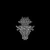 28562MC 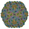 8eldC  8em6C M: map data used to model this data C: citing same article ( |
|---|---|
| Similar structure data |

-Links
 PDBj
PDBj- Assembly
| Deposited unit | 
|
|---|---|
| 1 | x 6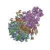
|
| 2 |
|
| 3 | 
|
| Symmetry | Point symmetry: (Schoenflies symbol: C6 (6 fold cyclic)) |
-Components
| #1: Protein | Mass: 56983.391 Da / Num. of mol.: 2 / Source method: isolated from a natural source / Source: (natural) Shigella phage Buco (virus) / References: UniProt: A0A482JG67#2: Protein | Mass: 20815.510 Da / Num. of mol.: 2 / Source method: isolated from a natural source / Source: (natural) Shigella phage Buco (virus) / References: UniProt: A0A482JKT9#3: Protein | | Mass: 84783.617 Da / Num. of mol.: 1 / Source method: isolated from a natural source / Source: (natural) Shigella phage Buco (virus) / References: UniProt: A0A482JLU9#4: Protein | Mass: 28595.883 Da / Num. of mol.: 3 / Source method: isolated from a natural source / Source: (natural) Shigella phage Buco (virus) / References: UniProt: A0A482JMG8 |
|---|
-Experimental details
-Experiment
| Experiment | Method: ELECTRON MICROSCOPY |
|---|---|
| EM experiment | Aggregation state: PARTICLE / 3D reconstruction method: single particle reconstruction |
- Sample preparation
| Component | Name: Shigella phage Buco / Type: VIRUS / Entity ID: all / Source: NATURAL |
|---|---|
| Source (natural) | Organism: Shigella phage Buco (virus) |
| Details of virus | Empty: NO / Enveloped: NO / Isolate: OTHER / Type: VIRION |
| Buffer solution | pH: 7.5 |
| Specimen | Embedding applied: NO / Shadowing applied: NO / Staining applied: NO / Vitrification applied: YES |
| Vitrification | Cryogen name: ETHANE |
- Electron microscopy imaging
| Experimental equipment |  Model: Titan Krios / Image courtesy: FEI Company |
|---|---|
| Microscopy | Model: FEI TITAN KRIOS |
| Electron gun | Electron source:  FIELD EMISSION GUN / Accelerating voltage: 300 kV / Illumination mode: FLOOD BEAM FIELD EMISSION GUN / Accelerating voltage: 300 kV / Illumination mode: FLOOD BEAM |
| Electron lens | Mode: BRIGHT FIELD / Nominal defocus max: 3000 nm / Nominal defocus min: 800 nm |
| Image recording | Electron dose: 33 e/Å2 / Film or detector model: GATAN K3 BIOQUANTUM (6k x 4k) |
- Processing
| CTF correction | Type: NONE | ||||||||||||||||||||||||
|---|---|---|---|---|---|---|---|---|---|---|---|---|---|---|---|---|---|---|---|---|---|---|---|---|---|
| 3D reconstruction | Resolution: 3.3 Å / Resolution method: FSC 0.143 CUT-OFF / Num. of particles: 24391 / Symmetry type: POINT | ||||||||||||||||||||||||
| Refine LS restraints |
|
