[English] 日本語
Yorodumi
- PDB-8erw: Precisely patterned nanofibers made from extendable protein multi... -

+
Open data


ID or keywords:

Loading...

-
Basic information

Entry
Database: PDB / ID: 8erw
TitlePrecisely patterned nanofibers made from extendable protein multiplexes
ComponentsC2HR4_8r
KeywordsDE NOVO PROTEIN / de novo design / nanofibers
Biological speciessynthetic construct (others)
MethodX-RAY DIFFRACTION / SYNCHROTRON / MOLECULAR REPLACEMENT / molecular replacement / Resolution: 2.88 Å
AuthorsBick, M.J. / Parmeggiani, F. / Bethel, N.P. / Sankaran, B. / Baker, D.
Funding support United States, 1items
OrganizationGrant numberCountry
Howard Hughes Medical Institute (HHMI) United States
Citation
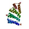
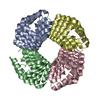
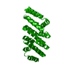
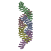
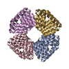
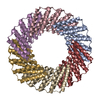
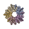
Journal: Nat Chem / Year: 2023
Title: Precisely patterned nanofibres made from extendable protein multiplexes.
Authors: Neville P Bethel / Andrew J Borst / Fabio Parmeggiani / Matthew J Bick / T J Brunette / Hannah Nguyen / Alex Kang / Asim K Bera / Lauren Carter / Marcos C Miranda / Ryan D Kibler / Mila Lamb ...Authors: Neville P Bethel / Andrew J Borst / Fabio Parmeggiani / Matthew J Bick / T J Brunette / Hannah Nguyen / Alex Kang / Asim K Bera / Lauren Carter / Marcos C Miranda / Ryan D Kibler / Mila Lamb / Xinting Li / Banumathi Sankaran / David Baker /
Abstract: Molecular systems with coincident cyclic and superhelical symmetry axes have considerable advantages for materials design as they can be readily lengthened or shortened by changing the length of the ...Molecular systems with coincident cyclic and superhelical symmetry axes have considerable advantages for materials design as they can be readily lengthened or shortened by changing the length of the constituent monomers. Among proteins, alpha-helical coiled coils have such symmetric, extendable architectures, but are limited by the relatively fixed geometry and flexibility of the helical protomers. Here we describe a systematic approach to generating modular and rigid repeat protein oligomers with coincident C to C and superhelical symmetry axes that can be readily extended by repeat propagation. From these building blocks, we demonstrate that a wide range of unbounded fibres can be systematically designed by introducing hydrophilic surface patches that force staggering of the monomers; the geometry of such fibres can be precisely tuned by varying the number of repeat units in the monomer and the placement of the hydrophilic patches.
History
DepositionOct 12, 2022Deposition site: RCSB / Processing site: RCSB
Revision 1.0Aug 23, 2023Provider: repository / Type: Initial release
Revision 1.1Sep 20, 2023Group: Database references / Category: citation / citation_author
Item: _citation.country / _citation.journal_abbrev ..._citation.country / _citation.journal_abbrev / _citation.journal_id_CSD / _citation.journal_id_ISSN / _citation.pdbx_database_id_DOI / _citation.pdbx_database_id_PubMed / _citation.title / _citation.year
Revision 1.2Dec 20, 2023Group: Database references / Category: citation / citation_author
Item: _citation.journal_volume / _citation.page_first ..._citation.journal_volume / _citation.page_first / _citation.page_last / _citation_author.identifier_ORCID

-
Structure visualization

Structure viewerMolecule:
MolmilJmol/JSmol

Downloads & links

-
Assembly

Deposited unit
A: C2HR4_8r
B: C2HR4_8r


Theoretical massNumber of molelcules
Total (without water)58,4942
Polymers58,4942
Non-polymers00
Water1629
1


  • Idetical with deposited unit
  • defined by author&software
  • Evidence: gel filtration
TypeNameSymmetry operationNumber
identity operation1_555x,y,z1
Buried area4220 Å2
ΔGint-45 kcal/mol
Surface area17670 Å2
MethodPISA
Unit cell
Length a, b, c (Å)55.318, 55.380, 145.134
Angle α, β, γ (deg.)90.000, 90.000, 90.000
Int Tables number19
Space group name H-MP212121

-
Components

#1: Protein C2HR4_8r


Mass: 29246.859 Da / Num. of mol.: 2
Source method: isolated from a genetically manipulated source
Source: (gene. exp.) synthetic construct (others) / Production host: Escherichia coli (E. coli)
#2: Water ChemComp-HOH / water


Mass: 18.015 Da / Num. of mol.: 9 / Source method: isolated from a natural source / Formula: H2O

-
Experimental details

-
Experiment

ExperimentMethod: X-RAY DIFFRACTION / Number of used crystals: 1

-
Sample preparation

CrystalDensity Matthews: 1.9 Å3/Da / Density % sol: 35.27 %
Crystal growTemperature: 293.15 K / Method: vapor diffusion, sitting drop / pH: 8.5
Details: 1:1 mixture of protein solution and crystallization solution consisting of 0.1M Tris-HCl pH 8.5, 25%(w/v) PEG 3000

-
Data collection

DiffractionMean temperature: 100 K / Ambient temp details: Liquid nitrogen vapor stream / Serial crystal experiment: N
Diffraction sourceSource: SYNCHROTRON / Site: ALS / Beamline: 8.2.2 / Wavelength: 1 Å
DetectorType: ADSC QUANTUM 315r / Detector: CCD / Date: Apr 5, 2016
RadiationMonochromator: Double-crystal Si(111) / Protocol: SINGLE WAVELENGTH / Monochromatic (M) / Laue (L): M / Scattering type: x-ray
Radiation wavelengthWavelength: 1 Å / Relative weight: 1
ReflectionResolution: 2.88→50 Å / Num. obs: 10228 / % possible obs: 96.9 % / Redundancy: 4.4 % / Biso Wilson estimate: 84.54 Å2 / Rpim(I) all: 0.055 / Rrim(I) all: 0.125 / Χ2: 0.927 / Net I/σ(I): 12.6
Reflection shellResolution: 2.88→2.94 Å / Redundancy: 3.4 % / Mean I/σ(I) obs: 1.18 / Num. unique obs: 134 / CC1/2: 0.667 / CC star: 0.894 / Rpim(I) all: 0.386 / Rrim(I) all: 0.776 / Χ2: 0.407 / % possible all: 89.2

-
Phasing

PhasingMethod: molecular replacement
Phasing MR
Highest resolutionLowest resolution
Rotation4.02 Å43.99 Å
Translation4.02 Å43.99 Å

-
Processing

Software
NameVersionClassification
HKL-20002.3.10data reduction
HKL-20002.3.10data scaling
PHASER2.7.18phasing
PHENIX1.19.1-4122-000refinement
PDB_EXTRACT3.27data extraction
RefinementMethod to determine structure: MOLECULAR REPLACEMENT
Starting model: Computational design model

Resolution: 2.88→43.99 Å / SU ML: 0.39 / Cross valid method: THROUGHOUT / σ(F): 1.33 / Phase error: 33.04 / Stereochemistry target values: ML
RfactorNum. reflection% reflection
Rfree0.2864 1028 10.14 %
Rwork0.2613 9108 -
obs0.2641 10136 95.67 %
Solvent computationShrinkage radii: 0.9 Å / VDW probe radii: 1.11 Å / Solvent model: FLAT BULK SOLVENT MODEL
Displacement parametersBiso max: 168.62 Å2 / Biso mean: 85.5266 Å2 / Biso min: 43.65 Å2
Refinement stepCycle: final / Resolution: 2.88→43.99 Å
ProteinNucleic acidLigandSolventTotal
Num. atoms3148 0 0 9 3157
Biso mean---71.98 -
Num. residues----509
LS refinement shell

Refine-ID: X-RAY DIFFRACTION / Rfactor Rfree error: 0 / Total num. of bins used: 7

Resolution (Å)Rfactor RfreeNum. reflection RfreeRfactor RworkNum. reflection RworkNum. reflection all% reflection obs (%)
2.88-3.030.33841380.30271189132790
3.03-3.220.30851460.26871316146299
3.22-3.470.43771450.33741294143997
3.47-3.820.34441430.29311296143997
3.82-4.370.24431530.22621304145796
4.37-5.510.25041510.24551352150397
5.51-43.990.2631520.25271357150993

+
About Yorodumi

-
News

-
Feb 9, 2022. New format data for meta-information of EMDB entries

New format data for meta-information of EMDB entries

  • Version 3 of the EMDB header file is now the official format.
  • The previous official version 1.9 will be removed from the archive.

Related info.:EMDB header

External links:wwPDB to switch to version 3 of the EMDB data model

-
Aug 12, 2020. Covid-19 info

Covid-19 info

URL: https://pdbj.org/emnavi/covid19.php

New page: Covid-19 featured information page in EM Navigator.

Related info.:Covid-19 info / Mar 5, 2020. Novel coronavirus structure data

+
Mar 5, 2020. Novel coronavirus structure data

Novel coronavirus structure data

Related info.:Yorodumi Speices / Aug 12, 2020. Covid-19 info

External links:COVID-19 featured content - PDBj / Molecule of the Month (242):Coronavirus Proteases

+
Jan 31, 2019. EMDB accession codes are about to change! (news from PDBe EMDB page)

EMDB accession codes are about to change! (news from PDBe EMDB page)

  • The allocation of 4 digits for EMDB accession codes will soon come to an end. Whilst these codes will remain in use, new EMDB accession codes will include an additional digit and will expand incrementally as the available range of codes is exhausted. The current 4-digit format prefixed with “EMD-” (i.e. EMD-XXXX) will advance to a 5-digit format (i.e. EMD-XXXXX), and so on. It is currently estimated that the 4-digit codes will be depleted around Spring 2019, at which point the 5-digit format will come into force.
  • The EM Navigator/Yorodumi systems omit the EMD- prefix.

Related info.:Q: What is EMD? / ID/Accession-code notation in Yorodumi/EM Navigator

External links:EMDB Accession Codes are Changing Soon! / Contact to PDBj

+
Jul 12, 2017. Major update of PDB

Major update of PDB

  • wwPDB released updated PDB data conforming to the new PDBx/mmCIF dictionary.
  • This is a major update changing the version number from 4 to 5, and with Remediation, in which all the entries are updated.
  • In this update, many items about electron microscopy experimental information are reorganized (e.g. em_software).
  • Now, EM Navigator and Yorodumi are based on the updated data.

External links:wwPDB Remediation / Enriched Model Files Conforming to OneDep Data Standards Now Available in the PDB FTP Archive

-
Yorodumi

Thousand views of thousand structures

  • Yorodumi is a browser for structure data from EMDB, PDB, SASBDB, etc.
  • This page is also the successor to EM Navigator detail page, and also detail information page/front-end page for Omokage search.
  • The word "yorodu" (or yorozu) is an old Japanese word meaning "ten thousand". "mi" (miru) is to see.

Related info.:EMDB / PDB / SASBDB / Comparison of 3 databanks / Yorodumi Search / Aug 31, 2016. New EM Navigator & Yorodumi / Yorodumi Papers / Jmol/JSmol / Function and homology information / Changes in new EM Navigator and Yorodumi

Read more