National Institutes of Health/National Institute Of Allergy and Infectious Diseases (NIH/NIAID)
米国
引用
ジャーナル: Blood / 年: 2023 タイトル: A bispecific inhibitor of complement and coagulation blocks activation in complementopathy models via a novel mechanism. 著者: John F Andersen / Haotian Lei / Ethan C Strayer / Tapan Kanai / Van Pham / Xiang-Zuo Pan / Patricia Hessab Alvarenga / Gloria F Gerber / Oluwatoyin A Asojo / Ivo M B Francischetti / Robert A ...著者: John F Andersen / Haotian Lei / Ethan C Strayer / Tapan Kanai / Van Pham / Xiang-Zuo Pan / Patricia Hessab Alvarenga / Gloria F Gerber / Oluwatoyin A Asojo / Ivo M B Francischetti / Robert A Brodsky / Jesus G Valenzuela / José M C Ribeiro / 要旨: Inhibitors of complement and coagulation are present in the saliva of a variety of blood-feeding arthropods that transmit parasitic and viral pathogens. Here, we describe the structure and mechanism ...Inhibitors of complement and coagulation are present in the saliva of a variety of blood-feeding arthropods that transmit parasitic and viral pathogens. Here, we describe the structure and mechanism of action of the sand fly salivary protein lufaxin, which inhibits the formation of the central alternative C3 convertase (C3bBb) and inhibits coagulation factor Xa (fXa). Surface plasmon resonance experiments show that lufaxin stabilizes the binding of serine protease factor B (FB) to C3b but does not detectably bind either C3b or FB alone. The crystal structure of the inhibitor reveals a novel all β-sheet fold containing 2 domains. A structure of the lufaxin-C3bB complex obtained via cryo-electron microscopy (EM) shows that lufaxin binds via its N-terminal domain at an interface containing elements of both C3b and FB. By occupying this spot, the inhibitor locks FB into a closed conformation in which proteolytic activation of FB by FD cannot occur. C3bB-bound lufaxin binds fXa at a separate site in its C-terminal domain. In the cryo-EM structure of a C3bB-lufaxin-fXa complex, the inhibitor binds to both targets simultaneously, and lufaxin inhibits fXa through substrate-like binding of a C-terminal peptide at the active site as well as other interactions in this region. Lufaxin inhibits complement activation in ex vivo models of atypical hemolytic uremic syndrome (aHUS) and paroxysmal nocturnal hemoglobinuria (PNH) as well as thrombin generation in plasma, providing a rationale for the development of a bispecific inhibitor to treat complement-related diseases in which thrombosis is a prominent manifestation.
 ムービー
ムービー コントローラー


 データを開く
 基本情報
基本情報 要素
要素 キーワード
キーワード 機能・相同性情報
 Lutzomyia longipalpis (昆虫)
 X線回折 /
  データ登録者
 米国, 1件
  引用
引用 構造の表示
構造の表示 ダウンロードとリンク
 その他のダウンロード
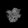


 PDBj
PDBj

 集合体
集合体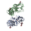


 Homo sapiens (ヒト) / 参照: UniProt: Q5WPU8
 タイプ: D-saccharide, beta linking / 分子量: 221.208 Da / 分子数: 2 / 由来タイプ: 合成 / 式: C8H15NO6


 分子量: 92.094 Da / 分子数: 1 / 由来タイプ: 合成 / 式: C3H8O3
 分子量: 79.904 Da / 分子数: 5 / 由来タイプ: 合成 / 式: Br
 試料調製
試料調製 解析
解析