 Movie
Movie Controller

[English] 日本語
 Yorodumi
- PDB-8enx: Crystal structure of beta'-COPI-WD40 domain Y33A mutant in comple... -
+ Open data

- Basic information
| Entry | Database: PDB / ID: 8enx | |||||||||
|---|---|---|---|---|---|---|---|---|---|---|
| Title | Crystal structure of beta'-COPI-WD40 domain Y33A mutant in complex with SARS-CoV-2 clientized spike tail heptapeptide. | |||||||||
 Components Components |
| |||||||||
 Keywords Keywords | PROTEIN TRANSPORT / COPI / protein trafficking / SARS-CoV-2 spike / dibasic motif | |||||||||
| Function / homology |  Function and homology information Function and homology informationCOPI vesicle coat / intra-Golgi vesicle-mediated transport / retrograde vesicle-mediated transport, Golgi to endoplasmic reticulum / endoplasmic reticulum to Golgi vesicle-mediated transport / intracellular protein transport / Golgi membrane / structural molecule activity Similarity search - Function | |||||||||
| Biological species |    Severe acute respiratory syndrome coronavirus 2 Severe acute respiratory syndrome coronavirus 2 | |||||||||
| Method |  X-RAY DIFFRACTION / SYNCHROTRON / MOLECULAR REPLACEMENT / Resolution: 1.8 Å X-RAY DIFFRACTION / SYNCHROTRON / MOLECULAR REPLACEMENT / Resolution: 1.8 Å | |||||||||
 Authors Authors | Dey, D. / Hasan, S.S. | |||||||||
| Funding support |  United States, 2items United States, 2items
| |||||||||
 Citation Citation | Journal: Nat Commun / Year: 2023 Title: A single C-terminal residue controls SARS-CoV-2 spike trafficking and incorporation into VLPs. Authors: Dey, D. / Qing, E. / He, Y. / Chen, Y. / Jennings, B. / Cohn, W. / Singh, S. / Gakhar, L. / Schnicker, N.J. / Pierce, B.G. / Whitelegge, J.P. / Doray, B. / Orban, J. / Gallagher, T. / Hasan, S.S. | |||||||||
| History |
|
- Structure visualization
| Structure viewer | Molecule: MolmilJmol/JSmol |
|---|
- Downloads & links
-Download
| PDBx/mmCIF format | 8enx.cif.gz | 312.9 KB | Display | PDBx/mmCIF format |
|---|---|---|---|---|
| PDB format | pdb8enx.ent.gz | 205.2 KB | Display | PDB format |
| PDBx/mmJSON format | 8enx.json.gz | Tree view | PDBx/mmJSON format | |
| Others |  Other downloads Other downloads |
-Validation report
| Arichive directory | https://data.pdbj.org/pub/pdb/validation_reports/en/8enxftp://data.pdbj.org/pub/pdb/validation_reports/en/8enx | HTTPS FTP |
|---|
-Related structure data
| Related structure data | 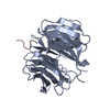 8ensC  8enwC  8enyC  8enzC 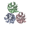 8eo0C  8szxC 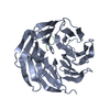 4j79S S: Starting model for refinement C: citing same article ( |
|---|---|
| Similar structure data |
-Links
 PDBj
PDBj

- Assembly
| Deposited unit | 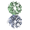
| ||||||||||||
|---|---|---|---|---|---|---|---|---|---|---|---|---|---|
| 1 | 
| ||||||||||||
| 2 | 
| ||||||||||||
| 3 | 
| ||||||||||||
| Unit cell |
|
-Components
| #1: Protein | Mass: 34288.633 Da / Num. of mol.: 2 / Mutation: Y33A Source method: isolated from a genetically manipulated source Source: (gene. exp.) Production host:  #2: Protein/peptide | | Mass: 846.970 Da / Num. of mol.: 1 / Source method: obtained synthetically Source: (synth.) Severe acute respiratory syndrome coronavirus 2#3: Water | ChemComp-HOH / |  Mass: 18.015 Da / Num. of mol.: 826 / Source method: isolated from a natural source / Formula: H2O Mass: 18.015 Da / Num. of mol.: 826 / Source method: isolated from a natural source / Formula: H2OHas protein modification | N | |
|---|
-Experimental details
-Experiment
| Experiment | Method: X-RAY DIFFRACTION / Number of used crystals: 1 |
|---|
- Sample preparation
| Crystal | Density Matthews: 2.21 Å3/Da / Density % sol: 44.35 % |
|---|---|
| Crystal grow | Temperature: 295.15 K / Method: vapor diffusion, hanging drop / pH: 6.2 / Details: 0.1M MES pH 6.2, 17% PEG20000 |
-Data collection
| Diffraction | Mean temperature: 100 K / Serial crystal experiment: N |
|---|---|
| Diffraction source | Source: SYNCHROTRON / Site: NSLS-II / Beamline: 17-ID-1 / Wavelength: 0.92 Å |
| Detector | Type: DECTRIS EIGER X 9M / Detector: PIXEL / Date: May 17, 2022 / Details: KB bimorph mirrors |
| Radiation | Monochromator: Si(111) DCM / Protocol: SINGLE WAVELENGTH / Monochromatic (M) / Laue (L): M / Scattering type: x-ray |
| Radiation wavelength | Wavelength: 0.92 Å / Relative weight: 1 |
| Reflection | Resolution: 1.8→28.33 Å / Num. obs: 53029 / % possible obs: 96.6 % / Redundancy: 2.4 % / Biso Wilson estimate: 16.09 Å2 / CC1/2: 0.989 / Rsym value: 0.137 / Net I/σ(I): 3.7 |
| Reflection shell | Resolution: 1.8→1.84 Å / Mean I/σ(I) obs: 1 / Num. unique obs: 2959 / CC1/2: 0.551 / Rsym value: 0.713 |
- Processing
| Software |
| ||||||||||||||||||||||||||||||||||||||||||||||||||||||||||||||||||||||||||||||||||||||||||||||||||||||||||||||||||||||||||||||||||||||||||||
|---|---|---|---|---|---|---|---|---|---|---|---|---|---|---|---|---|---|---|---|---|---|---|---|---|---|---|---|---|---|---|---|---|---|---|---|---|---|---|---|---|---|---|---|---|---|---|---|---|---|---|---|---|---|---|---|---|---|---|---|---|---|---|---|---|---|---|---|---|---|---|---|---|---|---|---|---|---|---|---|---|---|---|---|---|---|---|---|---|---|---|---|---|---|---|---|---|---|---|---|---|---|---|---|---|---|---|---|---|---|---|---|---|---|---|---|---|---|---|---|---|---|---|---|---|---|---|---|---|---|---|---|---|---|---|---|---|---|---|---|---|---|
| Refinement | Method to determine structure: MOLECULAR REPLACEMENT Starting model: 4J79 Resolution: 1.8→28.33 Å / SU ML: 0.2042 / Cross valid method: FREE R-VALUE / σ(F): 1.96 / Phase error: 18.0525 Stereochemistry target values: GeoStd + Monomer Library + CDL v1.2
| ||||||||||||||||||||||||||||||||||||||||||||||||||||||||||||||||||||||||||||||||||||||||||||||||||||||||||||||||||||||||||||||||||||||||||||
| Solvent computation | Shrinkage radii: 0.9 Å / VDW probe radii: 1.1 Å / Solvent model: FLAT BULK SOLVENT MODEL | ||||||||||||||||||||||||||||||||||||||||||||||||||||||||||||||||||||||||||||||||||||||||||||||||||||||||||||||||||||||||||||||||||||||||||||
| Displacement parameters | Biso mean: 19.23 Å2 | ||||||||||||||||||||||||||||||||||||||||||||||||||||||||||||||||||||||||||||||||||||||||||||||||||||||||||||||||||||||||||||||||||||||||||||
| Refinement step | Cycle: LAST / Resolution: 1.8→28.33 Å
| ||||||||||||||||||||||||||||||||||||||||||||||||||||||||||||||||||||||||||||||||||||||||||||||||||||||||||||||||||||||||||||||||||||||||||||
| Refine LS restraints |
| ||||||||||||||||||||||||||||||||||||||||||||||||||||||||||||||||||||||||||||||||||||||||||||||||||||||||||||||||||||||||||||||||||||||||||||
| LS refinement shell |
|
