beta-lactam antibiotic catabolic process / beta-lactamase activity / beta-lactamase / response to antibiotic Similarity search - Function
: / Beta-lactamase, class-A active site / Beta-lactamase class-A active site. / Beta-lactamase class A, catalytic domain / Beta-lactamase enzyme family / Beta-lactamase, class-A / Beta-lactamase/transpeptidase-like Similarity search - Domain/homology
Group: Advisory / Atomic model ...Advisory / Atomic model / Data collection / Database references / Derived calculations / Refinement description / Structure summary Category: atom_site / atom_site_anisotrop ...atom_site / atom_site_anisotrop / atom_type / entity / pdbx_contact_author / pdbx_database_related / pdbx_initial_refinement_model / pdbx_nonpoly_scheme / pdbx_struct_assembly / pdbx_validate_close_contact / pdbx_validate_torsion / refine / refine_hist / refine_ls_restr / refine_ls_shell / software / struct_conf / struct_conn / struct_mon_prot_cis Item: _entity.pdbx_number_of_molecules / _pdbx_struct_assembly.details ..._entity.pdbx_number_of_molecules / _pdbx_struct_assembly.details / _pdbx_struct_assembly.method_details / _refine.B_iso_mean / _refine.ls_R_factor_R_free / _refine.ls_R_factor_R_work / _refine.ls_R_factor_obs / _refine.ls_number_reflns_R_work / _refine.ls_number_reflns_obs / _refine.ls_percent_reflns_obs / _refine.overall_SU_ML / _refine.pdbx_overall_phase_error / _refine.pdbx_solvent_vdw_probe_radii / _refine_hist.number_atoms_solvent / _refine_hist.number_atoms_total / _refine_ls_restr.dev_ideal / _refine_ls_shell.R_factor_R_free / _refine_ls_shell.R_factor_R_work / _refine_ls_shell.number_reflns_R_work / _refine_ls_shell.percent_reflns_obs / _software.version / _struct_conn.pdbx_dist_value / _struct_mon_prot_cis.pdbx_omega_angle Description: Missing anisotropic B-factor Details: The actual reason for replacement was a request by one of the reviewers of the submitted manuscript requesting that, due to the high resolution of the structure, anisotropic B-factors and ...Details: The actual reason for replacement was a request by one of the reviewers of the submitted manuscript requesting that, due to the high resolution of the structure, anisotropic B-factors and hydrogen atoms have to be included in the structure. We agreed with that. A revised manuscript with the updated coordinates will be submitted soon for publication. Provider: author / Type: Coordinate replacement
In the structure databanks used in Yorodumi, some data are registered as the other names, "COVID-19 virus" and "2019-nCoV". Here are the details of the virus and the list of structure data.
Jan 31, 2019. EMDB accession codes are about to change! (news from PDBe EMDB page)
EMDB accession codes are about to change! (news from PDBe EMDB page)
The allocation of 4 digits for EMDB accession codes will soon come to an end. Whilst these codes will remain in use, new EMDB accession codes will include an additional digit and will expand incrementally as the available range of codes is exhausted. The current 4-digit format prefixed with “EMD-” (i.e. EMD-XXXX) will advance to a 5-digit format (i.e. EMD-XXXXX), and so on. It is currently estimated that the 4-digit codes will be depleted around Spring 2019, at which point the 5-digit format will come into force.
The EM Navigator/Yorodumi systems omit the EMD- prefix.
Related info.:Q: What is EMD? / ID/Accession-code notation in Yorodumi/EM Navigator
Yorodumi is a browser for structure data from EMDB, PDB, SASBDB, etc.
This page is also the successor to EM Navigator detail page, and also detail information page/front-end page for Omokage search.
The word "yorodu" (or yorozu) is an old Japanese word meaning "ten thousand". "mi" (miru) is to see.
Related info.:EMDB / PDB / SASBDB / Comparison of 3 databanks / Yorodumi Search / Aug 31, 2016. New EM Navigator & Yorodumi / Yorodumi Papers / Jmol/JSmol / Function and homology information / Changes in new EM Navigator and Yorodumi
 Movie
Movie Controller

 Yorodumi
 Open data
 Basic information
 Components
 Keywords
 Function and homology information
 Klebsiella pneumoniae (bacteria)
 X-RAY DIFFRACTION /
  Authors
 Argentina, 2items
  Citation
 Structure visualization
 Downloads & links
 Other downloads
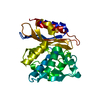
 PDBj
PDBj


 Assembly
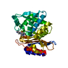
 Mass: 350.391 Da / Num. of mol.: 1 / Source method: obtained synthetically / Formula: C12H22N4O6S / Feature type: SUBJECT OF INVESTIGATION
 Mass: 18.015 Da / Num. of mol.: 297 / Source method: isolated from a natural source / Formula: H2O
 Sample preparation
 / Beamline: PROXIMA 2 / Wavelength: 0.885601 Å
 Processing