 Movie
Movie Controller

[English] 日本語
 Yorodumi
- PDB-8ed4: Structure of the complex between the arsenite oxidase and its nat... -
+ Open data

- Basic information
| Entry | Database: PDB / ID: 8ed4 | ||||||
|---|---|---|---|---|---|---|---|
| Title | Structure of the complex between the arsenite oxidase and its native electron acceptor cytochrome c552 from Pseudorhizobium sp. str. NT-26 | ||||||
 Components Components |
| ||||||
 Keywords Keywords | ELECTRON TRANSPORT / Complex | ||||||
| Function / homology |  Function and homology information Function and homology informationarsenate reductase (azurin) / oxidoreductase complex / molybdopterin cofactor binding / 3 iron, 4 sulfur cluster binding / NADH dehydrogenase activity / respiratory electron transport chain / 2 iron, 2 sulfur cluster binding / electron transfer activity / heme binding / membrane / metal ion binding Similarity search - Function | ||||||
| Biological species |  Pseudorhizobium banfieldiae (bacteria) Pseudorhizobium banfieldiae (bacteria) | ||||||
| Method |  X-RAY DIFFRACTION / SYNCHROTRON / MOLECULAR REPLACEMENT / Resolution: 2.25 Å X-RAY DIFFRACTION / SYNCHROTRON / MOLECULAR REPLACEMENT / Resolution: 2.25 Å | ||||||
 Authors Authors | Maher, M.J. / Poddar, N. | ||||||
| Funding support | 1items
| ||||||
 Citation Citation | Journal: Acta Crystallogr D Struct Biol / Year: 2023 Title: The structure of the complex between the arsenite oxidase from Pseudorhizobium banfieldiae sp. strain NT-26 and its native electron acceptor cytochrome c 552. Authors: Poddar, N. / Santini, J.M. / Maher, M.J. | ||||||
| History |
|
- Structure visualization
| Structure viewer | Molecule: MolmilJmol/JSmol |
|---|
- Downloads & links
-Download
| PDBx/mmCIF format | 8ed4.cif.gz | 873.3 KB | Display | PDBx/mmCIF format |
|---|---|---|---|---|
| PDB format | pdb8ed4.ent.gz | 698 KB | Display | PDB format |
| PDBx/mmJSON format | 8ed4.json.gz | Tree view | PDBx/mmJSON format | |
| Others |  Other downloads Other downloads |
-Validation report
| Arichive directory | https://data.pdbj.org/pub/pdb/validation_reports/ed/8ed4ftp://data.pdbj.org/pub/pdb/validation_reports/ed/8ed4 | HTTPS FTP |
|---|
-Related structure data


-Links
 PDBj
PDBj


- Assembly
| Deposited unit | 
| ||||||||
|---|---|---|---|---|---|---|---|---|---|
| 1 | 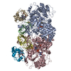
| ||||||||
| 2 | 
| ||||||||
| Unit cell |
|
-Components
-Protein , 3 types, 12 molecules ACEGBDFHIJKL
| #1: Protein | Mass: 93250.445 Da / Num. of mol.: 4 Source method: isolated from a genetically manipulated source Source: (gene. exp.) Pseudorhizobium banfieldiae (bacteria) / Gene: aroA, aioA, NT26_p10030 / Production host: #2: Protein | Mass: 17466.205 Da / Num. of mol.: 4 Source method: isolated from a genetically manipulated source Source: (gene. exp.) Pseudorhizobium banfieldiae (bacteria) / Gene: aroB / Production host: #3: Protein | Mass: 12736.475 Da / Num. of mol.: 4 Source method: isolated from a genetically manipulated source Source: (gene. exp.) Pseudorhizobium banfieldiae (bacteria) / Gene: cytC, NT26_p10031 / Production host: |
|---|
-Non-polymers , 10 types, 1780 molecules 


| #4: Chemical | ChemComp-MGD /  Mass: 740.557 Da / Num. of mol.: 8 / Source method: obtained synthetically / Formula: C20H26N10O13P2S2 Mass: 740.557 Da / Num. of mol.: 8 / Source method: obtained synthetically / Formula: C20H26N10O13P2S2#5: Chemical | ChemComp-4MO /  Mass: 95.940 Da / Num. of mol.: 4 / Source method: obtained synthetically / Formula: Mo Mass: 95.940 Da / Num. of mol.: 4 / Source method: obtained synthetically / Formula: Mo#6: Chemical | ChemComp-F3S /  Mass: 295.795 Da / Num. of mol.: 4 / Source method: obtained synthetically / Formula: Fe3S4 Mass: 295.795 Da / Num. of mol.: 4 / Source method: obtained synthetically / Formula: Fe3S4#7: Chemical | ChemComp-O /  Mass: 15.999 Da / Num. of mol.: 4 / Source method: obtained synthetically / Formula: O Mass: 15.999 Da / Num. of mol.: 4 / Source method: obtained synthetically / Formula: O#8: Chemical | ChemComp-FES /  Mass: 175.820 Da / Num. of mol.: 4 / Source method: obtained synthetically / Formula: Fe2S2 Mass: 175.820 Da / Num. of mol.: 4 / Source method: obtained synthetically / Formula: Fe2S2#9: Chemical |  Mass: 616.487 Da / Num. of mol.: 3 / Source method: obtained synthetically / Formula: C34H32FeN4O4 Mass: 616.487 Da / Num. of mol.: 3 / Source method: obtained synthetically / Formula: C34H32FeN4O4#10: Chemical | ChemComp-GOL / |  Mass: 92.094 Da / Num. of mol.: 1 / Source method: obtained synthetically / Formula: C3H8O3 Mass: 92.094 Da / Num. of mol.: 1 / Source method: obtained synthetically / Formula: C3H8O3#11: Chemical |  Mass: 194.226 Da / Num. of mol.: 2 / Source method: obtained synthetically / Formula: C8H18O5 / Comment: precipitant*YM Mass: 194.226 Da / Num. of mol.: 2 / Source method: obtained synthetically / Formula: C8H18O5 / Comment: precipitant*YM#12: Chemical | ChemComp-HEC / |  Mass: 618.503 Da / Num. of mol.: 1 / Source method: obtained synthetically / Formula: C34H34FeN4O4 Mass: 618.503 Da / Num. of mol.: 1 / Source method: obtained synthetically / Formula: C34H34FeN4O4#13: Water | ChemComp-HOH / | Mass: 18.015 Da / Num. of mol.: 1749 / Source method: isolated from a natural source / Formula: H2O |
|---|
-Details
| Has ligand of interest | N |
|---|---|
| Has protein modification | Y |
-Experimental details
-Experiment
| Experiment | Method: X-RAY DIFFRACTION / Number of used crystals: 1 |
|---|
- Sample preparation
| Crystal | Density Matthews: 2.3 Å3/Da / Density % sol: 46.59 % |
|---|---|
| Crystal grow | Temperature: 293 K / Method: vapor diffusion, hanging drop Details: 0.2 M sodium chloride, 0.1 M HEPES pH 7.3, 18% (w/v) PEG 3,350 |
-Data collection
| Diffraction | Mean temperature: 100 K / Serial crystal experiment: N |
|---|---|
| Diffraction source | Source: SYNCHROTRON / Site: Australian Synchrotron  / Beamline: MX2 / Wavelength: 0.953 Å / Beamline: MX2 / Wavelength: 0.953 Å |
| Detector | Type: DECTRIS EIGER X 16M / Detector: PIXEL / Date: Nov 4, 2020 |
| Radiation | Protocol: SINGLE WAVELENGTH / Monochromatic (M) / Laue (L): M / Scattering type: x-ray |
| Radiation wavelength | Wavelength: 0.953 Å / Relative weight: 1 |
| Reflection | Resolution: 2.25→49.23 Å / Num. obs: 214836 / % possible obs: 99.9 % / Redundancy: 7 % / CC1/2: 0.998 / Net I/σ(I): 11.4 |
| Reflection shell | Resolution: 2.25→2.29 Å / Num. unique obs: 10599 / CC1/2: 0.915 |
- Processing
| Software |
| ||||||||||||||||||||||||||||||||||||||||||||||||||||||||||||||||||||||||||||||||||||||||||||||||||||||||||||||||||||||||||||||||||||||||||||||||||||||||||||||||||||||||||||||||||||||
|---|---|---|---|---|---|---|---|---|---|---|---|---|---|---|---|---|---|---|---|---|---|---|---|---|---|---|---|---|---|---|---|---|---|---|---|---|---|---|---|---|---|---|---|---|---|---|---|---|---|---|---|---|---|---|---|---|---|---|---|---|---|---|---|---|---|---|---|---|---|---|---|---|---|---|---|---|---|---|---|---|---|---|---|---|---|---|---|---|---|---|---|---|---|---|---|---|---|---|---|---|---|---|---|---|---|---|---|---|---|---|---|---|---|---|---|---|---|---|---|---|---|---|---|---|---|---|---|---|---|---|---|---|---|---|---|---|---|---|---|---|---|---|---|---|---|---|---|---|---|---|---|---|---|---|---|---|---|---|---|---|---|---|---|---|---|---|---|---|---|---|---|---|---|---|---|---|---|---|---|---|---|---|---|
| Refinement | Method to determine structure: MOLECULAR REPLACEMENT Starting model: 4AAY,1CO6 Resolution: 2.25→49.23 Å / Cor.coef. Fo:Fc: 0.93 / Cor.coef. Fo:Fc free: 0.886 / SU B: 6.761 / SU ML: 0.163 / Cross valid method: THROUGHOUT / ESU R: 0.38 / ESU R Free: 0.236 / Stereochemistry target values: MAXIMUM LIKELIHOOD / Details: HYDROGENS HAVE BEEN ADDED IN THE RIDING POSITIONS
| ||||||||||||||||||||||||||||||||||||||||||||||||||||||||||||||||||||||||||||||||||||||||||||||||||||||||||||||||||||||||||||||||||||||||||||||||||||||||||||||||||||||||||||||||||||||
| Solvent computation | Ion probe radii: 0.8 Å / Shrinkage radii: 0.8 Å / VDW probe radii: 1.2 Å / Solvent model: BABINET MODEL WITH MASK | ||||||||||||||||||||||||||||||||||||||||||||||||||||||||||||||||||||||||||||||||||||||||||||||||||||||||||||||||||||||||||||||||||||||||||||||||||||||||||||||||||||||||||||||||||||||
| Displacement parameters | Biso mean: 11.397 Å2
| ||||||||||||||||||||||||||||||||||||||||||||||||||||||||||||||||||||||||||||||||||||||||||||||||||||||||||||||||||||||||||||||||||||||||||||||||||||||||||||||||||||||||||||||||||||||
| Refinement step | Cycle: 1 / Resolution: 2.25→49.23 Å
| ||||||||||||||||||||||||||||||||||||||||||||||||||||||||||||||||||||||||||||||||||||||||||||||||||||||||||||||||||||||||||||||||||||||||||||||||||||||||||||||||||||||||||||||||||||||
| Refine LS restraints |
|