 Movie
Movie Controller

[English] 日本語
 Yorodumi
- PDB-8ebo: Homopurine parallel G-quadruplex from human chromosome 7 stabiliz... -
+ Open data

- Basic information
| Entry | Database: PDB / ID: 8ebo | ||||||||||||||||||||||||||||
|---|---|---|---|---|---|---|---|---|---|---|---|---|---|---|---|---|---|---|---|---|---|---|---|---|---|---|---|---|---|
| Title | Homopurine parallel G-quadruplex from human chromosome 7 stabilized by K+ ions | ||||||||||||||||||||||||||||
 Components Components | DNA (5'-D(* Keywords KeywordsDNA / G-quadruplex / parallel / propeller loops | Function / homology | COBALT (III) ION / : / N-METHYLMESOPORPHYRIN / DNA / DNA (> 10) |  Function and homology information Function and homology informationBiological species |  Homo sapiens (human) Homo sapiens (human)Method |  X-RAY DIFFRACTION / SYNCHROTRON / MOLECULAR REPLACEMENT / Resolution: 1.98 Å X-RAY DIFFRACTION / SYNCHROTRON / MOLECULAR REPLACEMENT / Resolution: 1.98 Å  Authors AuthorsChen, E.V. / Yatsunyk, L.A. | Funding support | |  United States, 1items United States, 1items
 CitationJournal: Bioorg.Med.Chem. / Year: 2022 CitationJournal: Bioorg.Med.Chem. / Year: 2022Title: Homopurine guanine-rich sequences in complex with N-methyl mesoporphyrin IX form parallel G-quadruplex dimers and display a unique symmetry tetrad. Authors: Ye, M. / Chen, E.V. / Pfeil, S.H. / Martin, K.N. / Atrafi, T. / Yun, S. / Martinez, Z. / Yatsunyk, L.A. History |
|
- Structure visualization
| Structure viewer | Molecule: MolmilJmol/JSmol |
|---|
- Downloads & links
-Download
| PDBx/mmCIF format | 8ebo.cif.gz | 44.6 KB | Display | PDBx/mmCIF format |
|---|---|---|---|---|
| PDB format | pdb8ebo.ent.gz | 26.9 KB | Display | PDB format |
| PDBx/mmJSON format | 8ebo.json.gz | Tree view | PDBx/mmJSON format | |
| Others |  Other downloads Other downloads |
-Validation report
| Arichive directory | https://data.pdbj.org/pub/pdb/validation_reports/eb/8eboftp://data.pdbj.org/pub/pdb/validation_reports/eb/8ebo | HTTPS FTP |
|---|
-Related structure data
| Related structure data |  8edpC 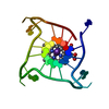 4g0fS S: Starting model for refinement C: citing same article ( |
|---|---|
| Similar structure data |
-Links
 PDBj
PDBj


- Assembly
| Deposited unit | 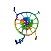
| ||||||||||||
|---|---|---|---|---|---|---|---|---|---|---|---|---|---|
| 1 |
| ||||||||||||
| Unit cell |
| ||||||||||||
| Components on special symmetry positions |
|
-Components
| #1: DNA chain | Mass: 7069.580 Da / Num. of mol.: 1 / Source method: obtained synthetically / Source: (synth.) Homo sapiens (human) | ||||||
|---|---|---|---|---|---|---|---|
| #2: Chemical | ChemComp-MMP /   Mass: 580.716 Da / Num. of mol.: 1 / Source method: obtained synthetically / Formula: C35H40N4O4 Mass: 580.716 Da / Num. of mol.: 1 / Source method: obtained synthetically / Formula: C35H40N4O4 | ||||||
| #3: Chemical |   Mass: 39.098 Da / Num. of mol.: 3 / Source method: obtained synthetically / Formula: K Mass: 39.098 Da / Num. of mol.: 3 / Source method: obtained synthetically / Formula: K#4: Chemical | ChemComp-3CO / |   Mass: 58.933 Da / Num. of mol.: 1 / Source method: obtained synthetically / Formula: Co Mass: 58.933 Da / Num. of mol.: 1 / Source method: obtained synthetically / Formula: Co#5: Water | ChemComp-HOH / |  Mass: 18.015 Da / Num. of mol.: 4 / Source method: isolated from a natural source / Formula: H2O Mass: 18.015 Da / Num. of mol.: 4 / Source method: isolated from a natural source / Formula: H2OHas ligand of interest | N | |
-Experimental details
-Experiment
| Experiment | Method: X-RAY DIFFRACTION / Number of used crystals: 1 |
|---|
- Sample preparation
| Crystal | Density Matthews: 2.18 Å3/Da / Density % sol: 43.6 % / Description: thin, well-ordered rhombuses |
|---|---|
| Crystal grow | Temperature: 285.15 K / Method: vapor diffusion, hanging drop Details: The well condition included 0.012 M NaCl, 0.08-0.1 M KCl, 0.04 M sodium cacodylate pH 5.5, 45% MPD, and 0.002 M hexammine cobalt(III) chloride |
-Data collection
| Diffraction | Mean temperature: 196 K / Serial crystal experiment: N |
|---|---|
| Diffraction source | Source: SYNCHROTRON / Site: APS / Beamline: 24-ID-C / Wavelength: 0.97918 Å |
| Detector | Type: DECTRIS EIGER X 16M / Detector: PIXEL / Date: Dec 8, 2021 |
| Radiation | Protocol: SINGLE WAVELENGTH / Monochromatic (M) / Laue (L): M / Scattering type: x-ray |
| Radiation wavelength | Wavelength: 0.97918 Å / Relative weight: 1 |
| Reflection | Resolution: 1.98→30.44 Å / Num. obs: 4296 / % possible obs: 99.2 % / Redundancy: 5.8 % / Biso Wilson estimate: 64.73 Å2 / CC1/2: 0.999 / Rmerge(I) obs: 0.049 / Rpim(I) all: 0.023 / Rrim(I) all: 0.054 / Net I/σ(I): 15.1 |
| Reflection shell | Resolution: 1.98→2.03 Å / Redundancy: 6.3 % / Mean I/σ(I) obs: 0.7 / Num. unique obs: 279 / CC1/2: 0.351 / Rpim(I) all: 0.894 / % possible all: 99.8 |
- Processing
| Software |
| ||||||||||||||||||||||||||||||||||||||||
|---|---|---|---|---|---|---|---|---|---|---|---|---|---|---|---|---|---|---|---|---|---|---|---|---|---|---|---|---|---|---|---|---|---|---|---|---|---|---|---|---|---|
| Refinement | Method to determine structure: MOLECULAR REPLACEMENT Starting model: 4G0F Resolution: 1.98→24.71 Å / SU ML: 0.2268 / Cross valid method: FREE R-VALUE / σ(F): 1.35 / Phase error: 45.9448 Stereochemistry target values: GeoStd + Monomer Library + CDL v1.2
| ||||||||||||||||||||||||||||||||||||||||
| Solvent computation | Shrinkage radii: 0.9 Å / VDW probe radii: 1.11 Å / Solvent model: FLAT BULK SOLVENT MODEL | ||||||||||||||||||||||||||||||||||||||||
| Displacement parameters | Biso mean: 95.26 Å2 | ||||||||||||||||||||||||||||||||||||||||
| Refinement step | Cycle: LAST / Resolution: 1.98→24.71 Å
| ||||||||||||||||||||||||||||||||||||||||
| Refine LS restraints |
| ||||||||||||||||||||||||||||||||||||||||
| LS refinement shell |
| ||||||||||||||||||||||||||||||||||||||||
| Refinement TLS params. | Method: refined / Origin x: -15.0484741614 Å / Origin y: 2.8269384339 Å / Origin z: -14.539514764 Å
| ||||||||||||||||||||||||||||||||||||||||
| Refinement TLS group | Selection details: all |
