 Movie
Movie Controller


+ Open data

- Basic information
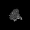
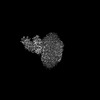
| Entry | Database: PDB / ID: 8eau | ||||||||||||||||||||||||||||||||||||||||||||||||||||||
|---|---|---|---|---|---|---|---|---|---|---|---|---|---|---|---|---|---|---|---|---|---|---|---|---|---|---|---|---|---|---|---|---|---|---|---|---|---|---|---|---|---|---|---|---|---|---|---|---|---|---|---|---|---|---|---|
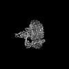
| Title | Yeast VO in complex with Vma21p | ||||||||||||||||||||||||||||||||||||||||||||||||||||||
 Components Components |
| ||||||||||||||||||||||||||||||||||||||||||||||||||||||
 Keywords Keywords | MEMBRANE PROTEIN / V-type / ATPase / assembly / proton | ||||||||||||||||||||||||||||||||||||||||||||||||||||||
| Function / homology |  Function and homology information Function and homology informationvacuolar proton-transporting V-type ATPase complex assembly / cell wall mannoprotein biosynthetic process / ATPase-coupled ion transmembrane transporter activity / protein localization to vacuolar membrane / cellular response to alkaline pH / Insulin receptor recycling / Transferrin endocytosis and recycling / polyphosphate metabolic process / ROS and RNS production in phagocytes / Golgi lumen acidification ...vacuolar proton-transporting V-type ATPase complex assembly / cell wall mannoprotein biosynthetic process / ATPase-coupled ion transmembrane transporter activity / protein localization to vacuolar membrane / cellular response to alkaline pH / Insulin receptor recycling / Transferrin endocytosis and recycling / polyphosphate metabolic process / ROS and RNS production in phagocytes / Golgi lumen acidification / Amino acids regulate mTORC1 / P-type proton-exporting transporter activity / vacuolar transport / vacuolar proton-transporting V-type ATPase, V0 domain / endosomal lumen acidification / vacuole organization / proton-transporting V-type ATPase complex / fungal-type vacuole / protein targeting to vacuole / vacuolar proton-transporting V-type ATPase complex / cellular hyperosmotic response / vacuolar acidification / fungal-type vacuole membrane / phosphatidylinositol-3,5-bisphosphate binding / proton transmembrane transporter activity / proton-transporting ATPase activity, rotational mechanism / intracellular copper ion homeostasis / Neutrophil degranulation / RNA endonuclease activity / endoplasmic reticulum-Golgi intermediate compartment membrane / proton transmembrane transport / cell periphery / ER to Golgi transport vesicle membrane / transmembrane transport / endocytosis / ATPase binding / protein-containing complex assembly / intracellular iron ion homeostasis / membrane raft / Golgi membrane / endoplasmic reticulum membrane / endoplasmic reticulum / membrane Similarity search - Function | ||||||||||||||||||||||||||||||||||||||||||||||||||||||
| Biological species |  | ||||||||||||||||||||||||||||||||||||||||||||||||||||||
| Method | ELECTRON MICROSCOPY / single particle reconstruction / cryo EM / Resolution: 3.1 Å | ||||||||||||||||||||||||||||||||||||||||||||||||||||||
 Authors Authors | Wang, H. / Bueler, S.A. / Rubinstein, J.L. | ||||||||||||||||||||||||||||||||||||||||||||||||||||||
| Funding support |  Canada, 1items Canada, 1items
| ||||||||||||||||||||||||||||||||||||||||||||||||||||||
 Citation Citation | Journal: Proc Natl Acad Sci U S A / Year: 2023 Title: Structural basis of V-ATPase V region assembly by Vma12p, 21p, and 22p. Authors: Hanlin Wang / Stephanie A Bueler / John L Rubinstein / Abstract: Vacuolar-type adenosine triphosphatases (V-ATPases) are rotary proton pumps that acidify specific intracellular compartments in almost all eukaryotic cells. These multi-subunit enzymes consist of a ...Vacuolar-type adenosine triphosphatases (V-ATPases) are rotary proton pumps that acidify specific intracellular compartments in almost all eukaryotic cells. These multi-subunit enzymes consist of a soluble catalytic V region and a membrane-embedded proton-translocating V region. V is assembled in the endoplasmic reticulum (ER) membrane, and V is assembled in the cytosol. However, V binds V only after V is transported to the Golgi membrane, thereby preventing acidification of the ER. We isolated V complexes and subcomplexes from bound to V-ATPase assembly factors Vma12p, Vma21p, and Vma22p. Electron cryomicroscopy shows how the Vma12-22p complex recruits subunits a, e, and f to the rotor ring of V while blocking premature binding of V. Vma21p, which contains an ER-retrieval motif, binds the V:Vma12-22p complex, "mature" V, and a complex that appears to contain a ring of loosely packed rotor subunits and the proteins YAR027W and YAR028W. The structures suggest that Vma21p binds assembly intermediates that contain a rotor ring and that activation of proton pumping following assembly of V with V removes Vma21p, allowing V-ATPase to remain in the Golgi. Together, these structures show how Vma12-22p and Vma21p function in V-ATPase assembly and quality control, ensuring the enzyme acidifies only its intended cellular targets. #1: Journal: Biorxiv / Year: 2022Title: Structural basis of V-ATPase V0 region assembly by Vma12p, 21p, and 22p Authors: Wang, H. / Bueler, S.A. / Rubinstein, J.L. | ||||||||||||||||||||||||||||||||||||||||||||||||||||||
| History |
|
- Structure visualization
| Structure viewer | Molecule: MolmilJmol/JSmol |
|---|
- Downloads & links
-Download
| PDBx/mmCIF format | 8eau.cif.gz | 487.9 KB | Display | PDBx/mmCIF format |
|---|---|---|---|---|
| PDB format | pdb8eau.ent.gz | 394.9 KB | Display | PDB format |
| PDBx/mmJSON format | 8eau.json.gz | Tree view | PDBx/mmJSON format | |
| Others |  Other downloads Other downloads |
-Validation report
| Arichive directory | https://data.pdbj.org/pub/pdb/validation_reports/ea/8eauftp://data.pdbj.org/pub/pdb/validation_reports/ea/8eau | HTTPS FTP |
|---|
-Related structure data
| Related structure data | 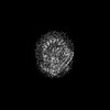 27986MC 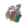 8easC  8eatC  8eavC M: map data used to model this data C: citing same article ( |
|---|---|
| Similar structure data |
-Links
 PDBj
PDBj


- Assembly
| Deposited unit | 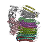
|
|---|---|
| 1 |
|
-Components
-V-type proton ATPase subunit ... , 6 types, 13 molecules acdeghijklmno
| #1: Protein | Mass: 95625.484 Da / Num. of mol.: 1 / Source method: isolated from a natural source / Source: (natural) | ||
|---|---|---|---|
| #3: Protein | Mass: 22610.641 Da / Num. of mol.: 1 / Source method: isolated from a natural source / Source: (natural) | ||
| #4: Protein | Mass: 39822.484 Da / Num. of mol.: 1 / Source method: isolated from a natural source / Source: (natural) | ||
| #5: Protein | Mass: 8387.065 Da / Num. of mol.: 1 / Source method: isolated from a natural source / Source: (natural) | ||
| #7: Protein | Mass: 16357.501 Da / Num. of mol.: 8 / Source method: isolated from a natural source / Source: (natural) #8: Protein | | Mass: 17046.361 Da / Num. of mol.: 1 / Source method: isolated from a natural source / Source: (natural) |
-Protein , 3 types, 3 molecules bfp
| #2: Protein | Mass: 29694.885 Da / Num. of mol.: 1 / Source method: isolated from a natural source / Source: (natural) |
|---|---|
| #6: Protein | Mass: 9369.934 Da / Num. of mol.: 1 / Source method: isolated from a natural source / Source: (natural) |
| #9: Protein | Mass: 8441.960 Da / Num. of mol.: 1 / Source method: isolated from a natural source / Source: (natural) |
-Details
| Has protein modification | N |
|---|
-Experimental details
-Experiment
| Experiment | Method: ELECTRON MICROSCOPY |
|---|---|
| EM experiment | Aggregation state: PARTICLE / 3D reconstruction method: single particle reconstruction |
- Sample preparation
| Component | Name: Yeast VO in complex with Vma21p / Type: COMPLEX / Entity ID: all / Source: NATURAL |
|---|---|
| Source (natural) | Organism: |
| Buffer solution | pH: 7.4 |
| Specimen | Embedding applied: NO / Shadowing applied: NO / Staining applied: NO / Vitrification applied: YES |
| Specimen support | Grid material: COPPER/RHODIUM / Grid type: Homemade |
| Vitrification | Instrument: LEICA EM GP / Cryogen name: ETHANE / Humidity: 80 % / Chamber temperature: 277 K |
- Electron microscopy imaging
| Experimental equipment |  Model: Titan Krios / Image courtesy: FEI Company |
|---|---|
| Microscopy | Model: FEI TITAN KRIOS |
| Electron gun | Electron source:  FIELD EMISSION GUN / Accelerating voltage: 300 kV / Illumination mode: FLOOD BEAM FIELD EMISSION GUN / Accelerating voltage: 300 kV / Illumination mode: FLOOD BEAM |
| Electron lens | Mode: BRIGHT FIELD / Nominal defocus max: 2000 nm / Nominal defocus min: 500 nm |
| Image recording | Electron dose: 49 e/Å2 / Film or detector model: FEI FALCON IV (4k x 4k) |
- Processing
| Software | Name: PHENIX / Version: 1.19.2_4158: / Classification: refinement | ||||||||||||||||||||||||
|---|---|---|---|---|---|---|---|---|---|---|---|---|---|---|---|---|---|---|---|---|---|---|---|---|---|
| EM software | Name: PHENIX / Category: model refinement | ||||||||||||||||||||||||
| CTF correction | Type: PHASE FLIPPING AND AMPLITUDE CORRECTION | ||||||||||||||||||||||||
| 3D reconstruction | Resolution: 3.1 Å / Resolution method: FSC 0.143 CUT-OFF / Num. of particles: 127922 / Symmetry type: POINT | ||||||||||||||||||||||||
| Refine LS restraints |
|
