 Movie
Movie Controller

[English] 日本語
 Yorodumi
- PDB-8eap: Cryo-EM structure of the in-situ gp10-gp26 from bacteriophage P22 -
+ Open data

- Basic information
| Entry | Database: PDB / ID: 8eap | ||||||
|---|---|---|---|---|---|---|---|
| Title | Cryo-EM structure of the in-situ gp10-gp26 from bacteriophage P22 | ||||||
 Components Components |
| ||||||
 Keywords Keywords | VIRAL PROTEIN / Bacteriophage P22 | ||||||
| Function / homology |  Function and homology information Function and homology informationsymbiont genome ejection through host cell envelope, short tail mechanism / virus tail / symbiont genome entry into host cell via pore formation in plasma membrane Similarity search - Function | ||||||
| Biological species |  Salmonella phage P22 (virus) Salmonella phage P22 (virus) | ||||||
| Method | ELECTRON MICROSCOPY / single particle reconstruction / cryo EM / Resolution: 3.3 Å | ||||||
 Authors Authors | Wang, C. / Liu, J. / Molineux, I.J. | ||||||
| Funding support |  United States, 1items United States, 1items
| ||||||
 Citation Citation | Journal: To Be Published Title: In-situ structure of tail machine reveals mechanistic insights into P22 assembly Authors: Wang, C. / Liu, J. / Molineux, I.J. | ||||||
| History |
|
- Structure visualization
| Structure viewer | Molecule: MolmilJmol/JSmol |
|---|
- Downloads & links
-Download
| PDBx/mmCIF format | 8eap.cif.gz | 503.2 KB | Display | PDBx/mmCIF format |
|---|---|---|---|---|
| PDB format | pdb8eap.ent.gz | 419.8 KB | Display | PDB format |
| PDBx/mmJSON format | 8eap.json.gz | Tree view | PDBx/mmJSON format | |
| Others |  Other downloads Other downloads |
-Validation report
| Arichive directory | https://data.pdbj.org/pub/pdb/validation_reports/ea/8eapftp://data.pdbj.org/pub/pdb/validation_reports/ea/8eap | HTTPS FTP |
|---|
-Related structure data
| Related structure data |  27790MC  8eb7C M: map data used to model this data C: citing same article ( |
|---|---|
| Similar structure data |


-Links
 PDBj
PDBj- Assembly
| Deposited unit | 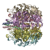
|
|---|---|
| 1 |
|
-Components
| #1: Protein | Mass: 6524.245 Da / Num. of mol.: 3 / Source method: isolated from a natural source / Source: (natural) Salmonella phage P22 (virus) / References: UniProt: P35837#2: Protein | Mass: 52410.852 Da / Num. of mol.: 6 / Source method: isolated from a natural source / Source: (natural) Salmonella phage P22 (virus) / References: UniProt: P26749Has protein modification | N | |
|---|
-Experimental details
-Experiment
| Experiment | Method: ELECTRON MICROSCOPY |
|---|---|
| EM experiment | Aggregation state: PARTICLE / 3D reconstruction method: single particle reconstruction |
- Sample preparation
| Component | Name: Cryo-EM structure of the in-situ gp10-gp26 from bacteriophage P22 Type: COMPLEX / Entity ID: all / Source: NATURAL |
|---|---|
| Source (natural) | Organism: Salmonella phage P22 (virus) |
| Buffer solution | pH: 7.5 |
| Specimen | Embedding applied: NO / Shadowing applied: NO / Staining applied: NO / Vitrification applied: YES |
| Vitrification | Cryogen name: ETHANE |
- Electron microscopy imaging
| Experimental equipment |  Model: Titan Krios / Image courtesy: FEI Company |
|---|---|
| Microscopy | Model: FEI TITAN KRIOS |
| Electron gun | Electron source:  FIELD EMISSION GUN / Accelerating voltage: 300 kV / Illumination mode: FLOOD BEAM FIELD EMISSION GUN / Accelerating voltage: 300 kV / Illumination mode: FLOOD BEAM |
| Electron lens | Mode: BRIGHT FIELD / Nominal defocus max: 1200 nm / Nominal defocus min: 600 nm |
| Image recording | Electron dose: 30 e/Å2 / Film or detector model: GATAN K3 (6k x 4k) |
- Processing
| Software | Name: PHENIX / Version: 1.19.2_4158: / Classification: refinement | ||||||||||||||||||||||||
|---|---|---|---|---|---|---|---|---|---|---|---|---|---|---|---|---|---|---|---|---|---|---|---|---|---|
| EM software | Name: PHENIX / Category: model refinement | ||||||||||||||||||||||||
| CTF correction | Type: PHASE FLIPPING AND AMPLITUDE CORRECTION | ||||||||||||||||||||||||
| 3D reconstruction | Resolution: 3.3 Å / Resolution method: FSC 0.143 CUT-OFF / Num. of particles: 106070 / Symmetry type: POINT | ||||||||||||||||||||||||
| Refine LS restraints |
|
