 Movie
Movie Controller

[English] 日本語
 Yorodumi
- PDB-8e9e: Rat protein farnesyltransferase in complex with FPP and inhibitor 2f -
+ Open data

- Basic information
| Entry | Database: PDB / ID: 8e9e | ||||||
|---|---|---|---|---|---|---|---|
| Title | Rat protein farnesyltransferase in complex with FPP and inhibitor 2f | ||||||
 Components Components |
| ||||||
 Keywords Keywords | TRANSFERASE/TRANSFERASE INHIBITOR / Inhibitor / Protein prenylyltransferase / Antifungal / TRANSFERASE / TRANSFERASE-Inhibitor complex / TRANSFERASE-TRANSFERASE INHIBITOR complex | ||||||
| Function / homology |  Function and homology information Function and homology informationApoptotic cleavage of cellular proteins / Inactivation, recovery and regulation of the phototransduction cascade / RAS processing / protein geranylgeranyltransferase activity / peptide pheromone maturation / protein geranylgeranyltransferase type I / CAAX-protein geranylgeranyltransferase activity / CAAX-protein geranylgeranyltransferase complex / protein farnesylation / protein farnesyltransferase ...Apoptotic cleavage of cellular proteins / Inactivation, recovery and regulation of the phototransduction cascade / RAS processing / protein geranylgeranyltransferase activity / peptide pheromone maturation / protein geranylgeranyltransferase type I / CAAX-protein geranylgeranyltransferase activity / CAAX-protein geranylgeranyltransferase complex / protein farnesylation / protein farnesyltransferase / protein farnesyltransferase activity / protein farnesyltransferase complex / Rab geranylgeranyltransferase activity / protein geranylgeranylation / regulation of fibroblast proliferation / positive regulation of skeletal muscle acetylcholine-gated channel clustering / nuclear envelope organization / geranylgeranyl diphosphate synthase activity / acetyltransferase activator activity / microtubule associated complex / enzyme-linked receptor protein signaling pathway / regulation of microtubule-based movement / alpha-tubulin binding / positive regulation of Rac protein signal transduction / positive regulation of cell cycle / lipid metabolic process / wound healing / protein maturation / receptor tyrosine kinase binding / positive regulation of fibroblast proliferation / microtubule binding / molecular adaptor activity / negative regulation of cell population proliferation / positive regulation of cell population proliferation / negative regulation of apoptotic process / enzyme binding / zinc ion binding / cytoplasm Similarity search - Function | ||||||
| Biological species |  | ||||||
| Method |  X-RAY DIFFRACTION / SYNCHROTRON / MOLECULAR REPLACEMENT / Resolution: 2.844 Å X-RAY DIFFRACTION / SYNCHROTRON / MOLECULAR REPLACEMENT / Resolution: 2.844 Å | ||||||
 Authors Authors | Wang, Y. / Shi, Y. / Beese, L.S. | ||||||
| Funding support |  United States, 1items United States, 1items
| ||||||
 Citation Citation | Journal: J.Med.Chem. / Year: 2022 Title: Structure-Guided Discovery of Potent Antifungals that Prevent Ras Signaling by Inhibiting Protein Farnesyltransferase. Authors: Wang, Y. / Xu, F. / Nichols, C.B. / Shi, Y. / Hellinga, H.W. / Alspaugh, J.A. / Distefano, M.D. / Beese, L.S. | ||||||
| History |
|
- Structure visualization
| Structure viewer | Molecule: MolmilJmol/JSmol |
|---|
- Downloads & links
-Download
| PDBx/mmCIF format | 8e9e.cif.gz | 170.4 KB | Display | PDBx/mmCIF format |
|---|---|---|---|---|
| PDB format | pdb8e9e.ent.gz | 130.3 KB | Display | PDB format |
| PDBx/mmJSON format | 8e9e.json.gz | Tree view | PDBx/mmJSON format | |
| Others |  Other downloads Other downloads |
-Validation report
| Arichive directory | https://data.pdbj.org/pub/pdb/validation_reports/e9/8e9eftp://data.pdbj.org/pub/pdb/validation_reports/e9/8e9e | HTTPS FTP |
|---|
-Related structure data
| Related structure data | 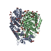 7t08C  7t09C  7t0aC  7t0bC  7t0cC  7t0dC  7t0eC  3e33S C: citing same article ( S: Starting model for refinement |
|---|---|
| Similar structure data |
-Links
 PDBj
PDBj

- Assembly
| Deposited unit | 
| ||||||||
|---|---|---|---|---|---|---|---|---|---|
| 1 |
| ||||||||
| Unit cell |
|
-Components
-Protein , 2 types, 2 molecules AB
| #1: Protein | Mass: 44098.145 Da / Num. of mol.: 1 Source method: isolated from a genetically manipulated source Source: (gene. exp.)  References: UniProt: Q04631, protein farnesyltransferase, protein geranylgeranyltransferase type I |
|---|---|
| #2: Protein | Mass: 48722.281 Da / Num. of mol.: 1 Source method: isolated from a genetically manipulated source Source: (gene. exp.) |
-Non-polymers , 6 types, 146 molecules 


| #3: Chemical | ChemComp-TRS /  Mass: 122.143 Da / Num. of mol.: 4 / Source method: obtained synthetically / Formula: C4H12NO3 / Comment: pH buffer*YM Mass: 122.143 Da / Num. of mol.: 4 / Source method: obtained synthetically / Formula: C4H12NO3 / Comment: pH buffer*YM#4: Chemical | ChemComp-EDO /  Mass: 62.068 Da / Num. of mol.: 9 / Source method: obtained synthetically / Formula: C2H6O2 Mass: 62.068 Da / Num. of mol.: 9 / Source method: obtained synthetically / Formula: C2H6O2#5: Chemical | ChemComp-FPP / |  Mass: 382.326 Da / Num. of mol.: 1 / Source method: obtained synthetically / Formula: C15H28O7P2 Mass: 382.326 Da / Num. of mol.: 1 / Source method: obtained synthetically / Formula: C15H28O7P2#6: Chemical | ChemComp-ZN / |  Mass: 65.409 Da / Num. of mol.: 1 / Source method: obtained synthetically / Formula: Zn Mass: 65.409 Da / Num. of mol.: 1 / Source method: obtained synthetically / Formula: Zn#7: Chemical | ChemComp-XMY / ( |  Mass: 565.425 Da / Num. of mol.: 1 / Source method: obtained synthetically / Formula: C26H28BrF3N4O2 / Feature type: SUBJECT OF INVESTIGATION Mass: 565.425 Da / Num. of mol.: 1 / Source method: obtained synthetically / Formula: C26H28BrF3N4O2 / Feature type: SUBJECT OF INVESTIGATION#8: Water | ChemComp-HOH / | Mass: 18.015 Da / Num. of mol.: 130 / Source method: isolated from a natural source / Formula: H2O |
|---|
-Details
| Has ligand of interest | Y |
|---|
-Experimental details
-Experiment
| Experiment | Method: X-RAY DIFFRACTION / Number of used crystals: 1 |
|---|
- Sample preparation
| Crystal | Density Matthews: 3.11 Å3/Da / Density % sol: 60.48 % |
|---|---|
| Crystal grow | Temperature: 290 K / Method: vapor diffusion, hanging drop / pH: 5.5 / Details: 50-150mM Na Acetate pH 5.5, 1-2.5% PEG8000 |
-Data collection
| Diffraction | Mean temperature: 100 K / Serial crystal experiment: N |
|---|---|
| Diffraction source | Source: SYNCHROTRON / Site: ALS / Beamline: 8.3.1 / Wavelength: 1.116 Å |
| Detector | Type: DECTRIS PILATUS3 S 6M / Detector: PIXEL / Date: May 10, 2019 |
| Radiation | Protocol: SINGLE WAVELENGTH / Monochromatic (M) / Laue (L): M / Scattering type: x-ray |
| Radiation wavelength | Wavelength: 1.116 Å / Relative weight: 1 |
| Reflection | Resolution: 2.844→43.38 Å / Num. obs: 26864 / % possible obs: 99 % / Redundancy: 5.8 % / CC1/2: 0.991 / CC star: 0.998 / Rmerge(I) obs: 0.1562 / Rpim(I) all: 0.0666 / Rrim(I) all: 0.1712 / Net I/σ(I): 10.49 |
| Reflection shell | Resolution: 2.844→2.946 Å / Redundancy: 5.4 % / Rmerge(I) obs: 0.7442 / Mean I/σ(I) obs: 1.98 / Num. unique obs: 2626 / CC1/2: 0.679 / CC star: 0.899 / Rpim(I) all: 0.3233 / Rrim(I) all: 0.8176 / % possible all: 97.8 |
- Processing
| Software |
| ||||||||||||||||||||||||||||||||||||||||||||||||||||||||||||||||||||||||||||||||||||||||||||||||||||||||||||||||||||||||||||||||||||||||||||||||||||||||||||||||||||||||
|---|---|---|---|---|---|---|---|---|---|---|---|---|---|---|---|---|---|---|---|---|---|---|---|---|---|---|---|---|---|---|---|---|---|---|---|---|---|---|---|---|---|---|---|---|---|---|---|---|---|---|---|---|---|---|---|---|---|---|---|---|---|---|---|---|---|---|---|---|---|---|---|---|---|---|---|---|---|---|---|---|---|---|---|---|---|---|---|---|---|---|---|---|---|---|---|---|---|---|---|---|---|---|---|---|---|---|---|---|---|---|---|---|---|---|---|---|---|---|---|---|---|---|---|---|---|---|---|---|---|---|---|---|---|---|---|---|---|---|---|---|---|---|---|---|---|---|---|---|---|---|---|---|---|---|---|---|---|---|---|---|---|---|---|---|---|---|---|---|---|
| Refinement | Method to determine structure: MOLECULAR REPLACEMENT / Starting model: 3.0E+33 / Resolution: 2.844→43.38 Å / SU ML: 0.32 / Cross valid method: THROUGHOUT / σ(F): 1.34 / Phase error: 18.81 / Stereochemistry target values: ML
| ||||||||||||||||||||||||||||||||||||||||||||||||||||||||||||||||||||||||||||||||||||||||||||||||||||||||||||||||||||||||||||||||||||||||||||||||||||||||||||||||||||||||
| Solvent computation | Shrinkage radii: 0.9 Å / VDW probe radii: 1.11 Å / Solvent model: FLAT BULK SOLVENT MODEL | ||||||||||||||||||||||||||||||||||||||||||||||||||||||||||||||||||||||||||||||||||||||||||||||||||||||||||||||||||||||||||||||||||||||||||||||||||||||||||||||||||||||||
| Displacement parameters | Biso max: 112.88 Å2 / Biso mean: 46.9063 Å2 / Biso min: 24.82 Å2 | ||||||||||||||||||||||||||||||||||||||||||||||||||||||||||||||||||||||||||||||||||||||||||||||||||||||||||||||||||||||||||||||||||||||||||||||||||||||||||||||||||||||||
| Refinement step | Cycle: final / Resolution: 2.844→43.38 Å
| ||||||||||||||||||||||||||||||||||||||||||||||||||||||||||||||||||||||||||||||||||||||||||||||||||||||||||||||||||||||||||||||||||||||||||||||||||||||||||||||||||||||||
| LS refinement shell | Refine-ID: X-RAY DIFFRACTION / Rfactor Rfree error: 0
|
