 Movie
Movie Controller

[English] 日本語
 Yorodumi
- PDB-8e75: Crystal structure of Pcryo_0616, the aminotransferase required to... -
+ Open data

- Basic information
| Entry | Database: PDB / ID: 8.0E+75 | ||||||
|---|---|---|---|---|---|---|---|
| Title | Crystal structure of Pcryo_0616, the aminotransferase required to synthesize UDP-N-acetyl-3-amino-D-glucosaminuronic acid (UDP-GlcNAc3NA) | ||||||
 Components Components | DegT/DnrJ/EryC1/StrS aminotransferase | ||||||
 Keywords Keywords | TRANSFERASE / aminotransferase / Psychrobacter cryohalolentis / carbohydratae | ||||||
| Function / homology | DegT/DnrJ/EryC1/StrS aminotransferase / DegT/DnrJ/EryC1/StrS aminotransferase family / polysaccharide biosynthetic process / transaminase activity / Pyridoxal phosphate-dependent transferase, small domain / Pyridoxal phosphate-dependent transferase, major domain / Pyridoxal phosphate-dependent transferase / pyridoxal phosphate binding / DegT/DnrJ/EryC1/StrS aminotransferase Function and homology information Function and homology information | ||||||
| Biological species |  Psychrobacter cryohalolentis K5 (bacteria) Psychrobacter cryohalolentis K5 (bacteria) | ||||||
| Method |  X-RAY DIFFRACTION / MOLECULAR REPLACEMENT / Resolution: 1.25 Å X-RAY DIFFRACTION / MOLECULAR REPLACEMENT / Resolution: 1.25 Å | ||||||
 Authors Authors | Hofmeister, D.L. / Seltzner, C.A. / Bockhaus, N.J. / thoden, J.B. / Holden, H.M. | ||||||
| Funding support |  United States, 1items United States, 1items
| ||||||
 Citation Citation | Journal: Protein Sci. / Year: 2023 Title: Investigation of the enzymes required for the biosynthesis of 2,3-diacetamido-2,3-dideoxy-d-glucuronic acid in Psychrobacter cryohalolentis K5 T. Authors: Hofmeister, D.L. / Seltzner, C.A. / Bockhaus, N.J. / Thoden, J.B. / Holden, H.M. | ||||||
| History |
|
- Structure visualization
| Structure viewer | Molecule: MolmilJmol/JSmol |
|---|
- Downloads & links
-Download
| PDBx/mmCIF format | 8e75.cif.gz | 108.4 KB | Display | PDBx/mmCIF format |
|---|---|---|---|---|
| PDB format | pdb8e75.ent.gz | 77.1 KB | Display | PDB format |
| PDBx/mmJSON format | 8e75.json.gz | Tree view | PDBx/mmJSON format | |
| Others |  Other downloads Other downloads |
-Validation report
| Arichive directory | https://data.pdbj.org/pub/pdb/validation_reports/e7/8e75ftp://data.pdbj.org/pub/pdb/validation_reports/e7/8e75 | HTTPS FTP |
|---|
-Related structure data
| Related structure data | 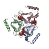 8e62C  8e77C  3nyuS S: Starting model for refinement C: citing same article ( |
|---|---|
| Similar structure data |
-Links
 PDBj
PDBj- Assembly
| Deposited unit | 
| ||||||||||||||||||||||||||||||||||||
|---|---|---|---|---|---|---|---|---|---|---|---|---|---|---|---|---|---|---|---|---|---|---|---|---|---|---|---|---|---|---|---|---|---|---|---|---|---|
| 1 | 
| ||||||||||||||||||||||||||||||||||||
| Unit cell |
| ||||||||||||||||||||||||||||||||||||
| Components on special symmetry positions |
|
-Components
| #1: Protein | Mass: 42248.543 Da / Num. of mol.: 1 Source method: isolated from a genetically manipulated source Source: (gene. exp.) Psychrobacter cryohalolentis K5 (bacteria)Strain: ATCC BAA-1226 / DSM 17306 / VKM B-2378 / K5 / Gene: Pcryo_0616 / Production host: | ||||
|---|---|---|---|---|---|
| #2: Chemical | ChemComp-NA /   Mass: 22.990 Da / Num. of mol.: 1 / Source method: obtained synthetically / Formula: Na Mass: 22.990 Da / Num. of mol.: 1 / Source method: obtained synthetically / Formula: Na | ||||
| #3: Chemical |   Mass: 62.068 Da / Num. of mol.: 3 / Source method: obtained synthetically / Formula: C2H6O2 Mass: 62.068 Da / Num. of mol.: 3 / Source method: obtained synthetically / Formula: C2H6O2#4: Water | ChemComp-HOH / |  Mass: 18.015 Da / Num. of mol.: 687 / Source method: isolated from a natural source / Formula: H2O Mass: 18.015 Da / Num. of mol.: 687 / Source method: isolated from a natural source / Formula: H2OHas ligand of interest | Y | |
-Experimental details
-Experiment
| Experiment | Method: X-RAY DIFFRACTION / Number of used crystals: 1 |
|---|
- Sample preparation
| Crystal | Density Matthews: 2.3 Å3/Da / Density % sol: 46.57 % |
|---|---|
| Crystal grow | Temperature: 293 K / Method: vapor diffusion, hanging drop / pH: 8 Details: Protein incubated with 5 mM UDP and 1 mM PLP. Precipitant: 18 - 22% poly(ethylene glycol) 8000, 200 mM LiCl, and 100 mM HEPPS (pH 8.0) |
-Data collection
| Diffraction | Mean temperature: 100 K / Serial crystal experiment: N |
|---|---|
| Diffraction source | Source: SEALED TUBE / Type: BRUKER D8 QUEST / Wavelength: 1.5418 Å |
| Detector | Type: Bruker PHOTON II / Detector: PIXEL / Date: Mar 2, 2019 |
| Radiation | Protocol: SINGLE WAVELENGTH / Monochromatic (M) / Laue (L): M / Scattering type: x-ray |
| Radiation wavelength | Wavelength: 1.5418 Å / Relative weight: 1 |
| Reflection | Resolution: 1.25→50 Å / Num. obs: 106460 / % possible obs: 97.8 % / Observed criterion σ(F): 0 / Observed criterion σ(I): 0 / Redundancy: 7.8 % / Rsym value: 0.051 / Net I/σ(I): 19.5 |
| Reflection shell | Resolution: 1.25→1.35 Å / Redundancy: 3.3 % / Mean I/σ(I) obs: 2.7 / Num. unique obs: 21148 / Rsym value: 0.42 / % possible all: 91.4 |
- Processing
| Software |
| ||||||||||||||||||||||||||||||||||||||||||||||||||||||||||||
|---|---|---|---|---|---|---|---|---|---|---|---|---|---|---|---|---|---|---|---|---|---|---|---|---|---|---|---|---|---|---|---|---|---|---|---|---|---|---|---|---|---|---|---|---|---|---|---|---|---|---|---|---|---|---|---|---|---|---|---|---|---|
| Refinement | Method to determine structure: MOLECULAR REPLACEMENT Starting model: 3nyu Resolution: 1.25→28.93 Å / Cor.coef. Fo:Fc: 0.973 / Cor.coef. Fo:Fc free: 0.969 / SU B: 0.738 / SU ML: 0.03 / Cross valid method: THROUGHOUT / σ(F): 0 / ESU R: 0.04 / ESU R Free: 0.041 / Stereochemistry target values: MAXIMUM LIKELIHOOD Details: HYDROGENS HAVE BEEN ADDED IN THE RIDING POSITIONS U VALUES : REFINED INDIVIDUALLY
| ||||||||||||||||||||||||||||||||||||||||||||||||||||||||||||
| Solvent computation | Ion probe radii: 0.8 Å / Shrinkage radii: 0.8 Å / VDW probe radii: 1.2 Å / Solvent model: MASK | ||||||||||||||||||||||||||||||||||||||||||||||||||||||||||||
| Displacement parameters | Biso max: 61.88 Å2 / Biso mean: 10.402 Å2 / Biso min: 3.81 Å2
| ||||||||||||||||||||||||||||||||||||||||||||||||||||||||||||
| Refinement step | Cycle: final / Resolution: 1.25→28.93 Å
| ||||||||||||||||||||||||||||||||||||||||||||||||||||||||||||
| Refine LS restraints |
| ||||||||||||||||||||||||||||||||||||||||||||||||||||||||||||
| LS refinement shell | Resolution: 1.25→1.278 Å / Rfactor Rfree error: 0
|
