 Movie
Movie Controller

[English] 日本語
 Yorodumi
- PDB-8e62: STRUCTURE OF Pcryo_0615 from Psychrobacter cryohalolentis, an N-a... -
+ Open data

- Basic information
| Entry | Database: PDB / ID: 8.0E+62 | ||||||
|---|---|---|---|---|---|---|---|
| Title | STRUCTURE OF Pcryo_0615 from Psychrobacter cryohalolentis, an N-acetyltransferase required to produce Diacetamido-2,3-dideoxy-D-glucuronic acid | ||||||
 Components Components | UDP-N-acetylglucosamine acyltransferase | ||||||
 Keywords Keywords | TRANSFERASE / N-acetyltransferase / 2 / 3-diacetamido-2 / 3-dideoxy-D-glucuronic acid / Psychrobacter cryohalolentis | ||||||
| Function / homology | acyl-[acyl-carrier-protein]-UDP-N-acetylglucosamine O-acyltransferase activity / Acyl-[acyl-carrier-protein]--UDP-N-acetylglucosamine O-acyltransferase / Hexapeptide repeat / Bacterial transferase hexapeptide (six repeats) / lipid A biosynthetic process / Trimeric LpxA-like superfamily / COENZYME A / Chem-MJZ / UDP-N-acetylglucosamine acyltransferase Function and homology information Function and homology information | ||||||
| Biological species |  Psychrobacter cryohalolentis K5 (bacteria) Psychrobacter cryohalolentis K5 (bacteria) | ||||||
| Method |  X-RAY DIFFRACTION / SYNCHROTRON / MOLECULAR REPLACEMENT / Resolution: 1.8 Å X-RAY DIFFRACTION / SYNCHROTRON / MOLECULAR REPLACEMENT / Resolution: 1.8 Å | ||||||
 Authors Authors | Hofmeister, D.L. / Bockhaus, N.J. / Seltzner, C.A. / Thoden, J.B. / Holden, H.M. | ||||||
| Funding support |  United States, 1items United States, 1items
| ||||||
 Citation Citation | Journal: Protein Sci. / Year: 2023 Title: Investigation of the enzymes required for the biosynthesis of 2,3-diacetamido-2,3-dideoxy-d-glucuronic acid in Psychrobacter cryohalolentis K5 T. Authors: Hofmeister, D.L. / Seltzner, C.A. / Bockhaus, N.J. / Thoden, J.B. / Holden, H.M. | ||||||
| History |
|
- Structure visualization
| Structure viewer | Molecule: MolmilJmol/JSmol |
|---|
- Downloads & links
-Download
| PDBx/mmCIF format | 8e62.cif.gz | 241.4 KB | Display | PDBx/mmCIF format |
|---|---|---|---|---|
| PDB format | pdb8e62.ent.gz | 191.7 KB | Display | PDB format |
| PDBx/mmJSON format | 8e62.json.gz | Tree view | PDBx/mmJSON format | |
| Others |  Other downloads Other downloads |
-Validation report
| Arichive directory | https://data.pdbj.org/pub/pdb/validation_reports/e6/8e62ftp://data.pdbj.org/pub/pdb/validation_reports/e6/8e62 | HTTPS FTP |
|---|
-Related structure data
| Related structure data |  8e75C  8e77C  5demS S: Starting model for refinement C: citing same article ( |
|---|---|
| Similar structure data |
-Links
 PDBj
PDBj


- Assembly
| Deposited unit | 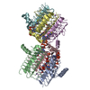
| ||||||||
|---|---|---|---|---|---|---|---|---|---|
| 1 | 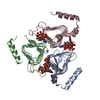
| ||||||||
| 2 | 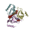
| ||||||||
| Unit cell |
|
-Components
| #1: Protein | Mass: 20516.600 Da / Num. of mol.: 6 Source method: isolated from a genetically manipulated source Source: (gene. exp.) Psychrobacter cryohalolentis K5 (bacteria)Strain: ATCC BAA-1226 / DSM 17306 / VKM B-2378 / K5 / Gene: Pcryo_0615 / Production host: #2: Chemical |   Mass: 620.352 Da / Num. of mol.: 3 / Source method: obtained synthetically / Formula: C17H26N4O17P2 / Feature type: SUBJECT OF INVESTIGATION Mass: 620.352 Da / Num. of mol.: 3 / Source method: obtained synthetically / Formula: C17H26N4O17P2 / Feature type: SUBJECT OF INVESTIGATION#3: Chemical | ChemComp-COA /   Mass: 767.534 Da / Num. of mol.: 6 / Source method: obtained synthetically / Formula: C21H36N7O16P3S / Feature type: SUBJECT OF INVESTIGATION Mass: 767.534 Da / Num. of mol.: 6 / Source method: obtained synthetically / Formula: C21H36N7O16P3S / Feature type: SUBJECT OF INVESTIGATION#4: Chemical |   Mass: 22.990 Da / Num. of mol.: 2 / Source method: obtained synthetically / Formula: Na Mass: 22.990 Da / Num. of mol.: 2 / Source method: obtained synthetically / Formula: Na#5: Water | ChemComp-HOH / |  Mass: 18.015 Da / Num. of mol.: 631 / Source method: isolated from a natural source / Formula: H2O Mass: 18.015 Da / Num. of mol.: 631 / Source method: isolated from a natural source / Formula: H2OHas ligand of interest | Y | |
|---|
-Experimental details
-Experiment
| Experiment | Method: X-RAY DIFFRACTION / Number of used crystals: 1 |
|---|
- Sample preparation
| Crystal | Density Matthews: 2.41 Å3/Da / Density % sol: 48.97 % |
|---|---|
| Crystal grow | Temperature: 293 K / Method: vapor diffusion, hanging drop / pH: 6 Details: Protein incubated with: 5 mM CoA and 5 mM UDP-GlcNAc3NA. Precipitant: 16 - 20% PEG-3350, 2% 2-methyl-2,4-pentanediol, and 100 mM MES (pH 6.0) |
-Data collection
| Diffraction | Mean temperature: 100 K / Serial crystal experiment: N |
|---|---|
| Diffraction source | Source: SYNCHROTRON / Site: APS / Beamline: 19-BM / Wavelength: 0.9792 Å |
| Detector | Type: ADSC QUANTUM 270 / Detector: CCD / Date: Aug 18, 2019 |
| Radiation | Protocol: SINGLE WAVELENGTH / Monochromatic (M) / Laue (L): M / Scattering type: x-ray |
| Radiation wavelength | Wavelength: 0.9792 Å / Relative weight: 1 |
| Reflection | Resolution: 1.8→50 Å / Num. obs: 102909 / % possible obs: 98.1 % / Observed criterion σ(F): 0 / Observed criterion σ(I): 0 / Redundancy: 4.3 % / Rsym value: 0.079 / Net I/σ(I): 30.8 |
| Reflection shell | Resolution: 1.8→1.83 Å / Redundancy: 3.6 % / Mean I/σ(I) obs: 5.3 / Num. unique obs: 5043 / Rsym value: 0.226 / % possible all: 96.2 |
- Processing
| Software |
| ||||||||||||||||||||||||||||||||||||||||||||||||||||||||||||
|---|---|---|---|---|---|---|---|---|---|---|---|---|---|---|---|---|---|---|---|---|---|---|---|---|---|---|---|---|---|---|---|---|---|---|---|---|---|---|---|---|---|---|---|---|---|---|---|---|---|---|---|---|---|---|---|---|---|---|---|---|---|
| Refinement | Method to determine structure: MOLECULAR REPLACEMENT Starting model: 5dem Resolution: 1.8→48.15 Å / Cor.coef. Fo:Fc: 0.961 / Cor.coef. Fo:Fc free: 0.937 / SU B: 2.297 / SU ML: 0.072 / Cross valid method: THROUGHOUT / σ(F): 0 / ESU R: 0.12 / ESU R Free: 0.118 / Stereochemistry target values: MAXIMUM LIKELIHOOD Details: HYDROGENS HAVE BEEN ADDED IN THE RIDING POSITIONS U VALUES : REFINED INDIVIDUALLY
| ||||||||||||||||||||||||||||||||||||||||||||||||||||||||||||
| Solvent computation | Ion probe radii: 0.8 Å / Shrinkage radii: 0.8 Å / VDW probe radii: 1.2 Å / Solvent model: MASK | ||||||||||||||||||||||||||||||||||||||||||||||||||||||||||||
| Displacement parameters | Biso max: 114.05 Å2 / Biso mean: 22.1 Å2 / Biso min: 9.31 Å2
| ||||||||||||||||||||||||||||||||||||||||||||||||||||||||||||
| Refinement step | Cycle: final / Resolution: 1.8→48.15 Å
| ||||||||||||||||||||||||||||||||||||||||||||||||||||||||||||
| Refine LS restraints |
| ||||||||||||||||||||||||||||||||||||||||||||||||||||||||||||
| LS refinement shell | Resolution: 1.802→1.849 Å / Rfactor Rfree error: 0 / Total num. of bins used: 20
|
