Movie
Movie Controller


+ Open data

- Basic information
| Entry | Database: PDB / ID: 8.0E+31 | ||||||
|---|---|---|---|---|---|---|---|
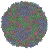
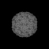
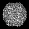
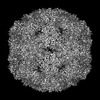
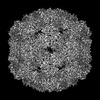
| Title | Purification of Enterovirus A71, strain 4643, WT capsid | ||||||
 Components Components |
| ||||||
 Keywords Keywords | VIRUS / enterovirus / thermostability / capsid | ||||||
| Function / homology |  Function and homology information Function and homology informationsymbiont genome entry into host cell via pore formation in plasma membrane / virion component / viral capsid / host cell / host cell cytoplasm / symbiont-mediated suppression of host gene expression / symbiont entry into host cell / virion attachment to host cell / structural molecule activity Similarity search - Function | ||||||
| Biological species |   Enterovirus A71 Enterovirus A71 | ||||||
| Method | ELECTRON MICROSCOPY / single particle reconstruction / cryo EM / Resolution: 14 Å | ||||||
 Authors Authors | Catching, A. / Capponi, S. / Andino, R. | ||||||
| Funding support |  United States, 1items United States, 1items
| ||||||
 Citation Citation | Journal: Nat Commun / Year: 2023 Title: A tradeoff between enterovirus A71 particle stability and cell entry. Authors: Adam Catching / Ming Te Yeh / Simone Bianco / Sara Capponi / Raul Andino / Abstract: A central role of viral capsids is to protect the viral genome from the harsh extracellular environment while facilitating initiation of infection when the virus encounters a target cell. Viruses are ...A central role of viral capsids is to protect the viral genome from the harsh extracellular environment while facilitating initiation of infection when the virus encounters a target cell. Viruses are thought to have evolved an optimal equilibrium between particle stability and efficiency of cell entry. In this study, we genetically perturb this equilibrium in a non-enveloped virus, enterovirus A71 to determine its structural basis. We isolate a single-point mutation variant with increased particle thermotolerance and decreased efficiency of cell entry. Using cryo-electron microscopy and molecular dynamics simulations, we determine that the thermostable native particles have acquired an expanded conformation that results in a significant increase in protein dynamics. Examining the intermediate states of the thermostable variant reveals a potential pathway for uncoating. We propose a sequential release of the lipid pocket factor, followed by internal VP4 and ultimately the viral RNA. | ||||||
| History |
|
- Structure visualization
| Structure viewer | Molecule: MolmilJmol/JSmol |
|---|
- Downloads & links
-Download
| PDBx/mmCIF format | 8e31.cif.gz | 129.6 KB | Display | PDBx/mmCIF format |
|---|---|---|---|---|
| PDB format | pdb8e31.ent.gz | 96.7 KB | Display | PDB format |
| PDBx/mmJSON format | 8e31.json.gz | Tree view | PDBx/mmJSON format | |
| Others |  Other downloads Other downloads |
-Validation report
| Summary document | 8e31_validation.pdf.gz | 1.3 MB | Display | wwPDB validaton report |
|---|---|---|---|---|
| Full document | 8e31_full_validation.pdf.gz | 1.3 MB | Display | |
| Data in XML | 8e31_validation.xml.gz | 36.3 KB | Display | |
| Data in CIF | 8e31_validation.cif.gz | 53.1 KB | Display | |
| Arichive directory | https://data.pdbj.org/pub/pdb/validation_reports/e3/8e31ftp://data.pdbj.org/pub/pdb/validation_reports/e3/8e31 | HTTPS FTP |
-Related structure data
| Related structure data | 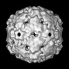 27853MC  8e2xC  8e2yC  8e38C 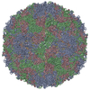 8e39C 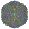 8e3aC 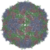 8e3bC  8e3cC M: map data used to model this data C: citing same article ( |
|---|---|
| Similar structure data |
-Links
 PDBj
PDBj
- Assembly
| Deposited unit | 
|
|---|---|
| 1 | x 60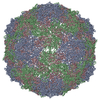
|
| 2 |
|
| 3 | x 5
|
| 4 | x 6
|
| 5 | 
|
| Symmetry | Point symmetry: (Schoenflies symbol: I (icosahedral)) |
-Components
| #1: Protein | Mass: 25366.764 Da / Num. of mol.: 1 Source method: isolated from a genetically manipulated source Source: (gene. exp.) Enterovirus A71 / Strain: Tainan/4643/98 / Cell line (production host): RD / Production host:  Homo sapiens (human) / Strain (production host): RD / Tissue (production host): Muscle / References: UniProt: F8SSP9 Homo sapiens (human) / Strain (production host): RD / Tissue (production host): Muscle / References: UniProt: F8SSP9 |
|---|---|
| #2: Protein | Mass: 24917.145 Da / Num. of mol.: 1 Source method: isolated from a genetically manipulated source Source: (gene. exp.) Enterovirus A71 / Cell line (production host): RD / Production host: Homo sapiens (human) / Strain (production host): RD / Tissue (production host): Muscle / References: UniProt: W8XW39 |
| #3: Protein | Mass: 23453.926 Da / Num. of mol.: 1 Source method: isolated from a genetically manipulated source Source: (gene. exp.) Enterovirus A71 / Strain: Tainan/4643/98 / Cell line (production host): RD / Production host: Homo sapiens (human) / Strain (production host): RD / Tissue (production host): Muscle / References: UniProt: W8XW58 |
-Experimental details
-Experiment
| Experiment | Method: ELECTRON MICROSCOPY |
|---|---|
| EM experiment | Aggregation state: PARTICLE / 3D reconstruction method: single particle reconstruction |
- Sample preparation
| Component | Name: Human enterovirus 71 / Type: VIRUS / Entity ID: all / Source: RECOMBINANT |
|---|---|
| Molecular weight | Experimental value: NO |
| Source (natural) | Organism: Human enterovirus 71 |
| Source (recombinant) | Organism: Homo sapiens (human) |
| Details of virus | Empty: NO / Enveloped: NO / Isolate: STRAIN / Type: VIRUS-LIKE PARTICLE |
| Natural host | Organism: Homo sapiens |
| Virus shell | Name: Capsid / Diameter: 300 nm / Triangulation number (T number): 1 |
| Buffer solution | pH: 7 |
| Specimen | Embedding applied: NO / Shadowing applied: NO / Staining applied: NO / Vitrification applied: YES |
| Specimen support | Grid material: COPPER / Grid mesh size: 200 divisions/in. / Grid type: Quantifoil R2/1 |
| Vitrification | Instrument: FEI VITROBOT MARK IV / Cryogen name: ETHANE / Humidity: 100 % / Chamber temperature: 297 K |
- Electron microscopy imaging
| Experimental equipment |  Model: Talos Arctica / Image courtesy: FEI Company |
|---|---|
| Microscopy | Model: FEI TECNAI ARCTICA |
| Electron gun | Electron source:  FIELD EMISSION GUN / Accelerating voltage: 200 kV / Illumination mode: FLOOD BEAM FIELD EMISSION GUN / Accelerating voltage: 200 kV / Illumination mode: FLOOD BEAM |
| Electron lens | Mode: BRIGHT FIELD / Nominal magnification: 45000 X / Nominal defocus max: 2000 nm / Nominal defocus min: 1500 nm / Cs: 2.7 mm |
| Specimen holder | Cryogen: NITROGEN |
| Image recording | Average exposure time: 6 sec. / Electron dose: 64.1 e/Å2 / Film or detector model: GATAN K3 (6k x 4k) / Num. of grids imaged: 1 |
| Image scans | Width: 6000 / Height: 4000 |
- Processing
| Software | Name: PHENIX / Version: 1.19.2_4158: / Classification: refinement | ||||||||||||||||||||||||||||
|---|---|---|---|---|---|---|---|---|---|---|---|---|---|---|---|---|---|---|---|---|---|---|---|---|---|---|---|---|---|
| EM software |
| ||||||||||||||||||||||||||||
| CTF correction | Type: PHASE FLIPPING ONLY | ||||||||||||||||||||||||||||
| Particle selection | Num. of particles selected: 20699 | ||||||||||||||||||||||||||||
| 3D reconstruction | Resolution: 14 Å / Resolution method: FSC 0.143 CUT-OFF / Num. of particles: 848 / Algorithm: BACK PROJECTION / Num. of class averages: 1 / Symmetry type: POINT | ||||||||||||||||||||||||||||
| Atomic model building | Protocol: RIGID BODY FIT / Space: REAL / Target criteria: Correlation Coefficient | ||||||||||||||||||||||||||||
| Atomic model building | PDB-ID: 3VBS Accession code: 3VBS / Source name: PDB / Type: experimental model | ||||||||||||||||||||||||||||
| Refine LS restraints |
|