 Movie
Movie Controller


+ Open data

- Basic information
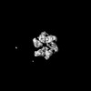
| Entry | Database: PDB / ID: 8e2l | |||||||||||||||||||||||||||||||||||||||||||||||||||||||||
|---|---|---|---|---|---|---|---|---|---|---|---|---|---|---|---|---|---|---|---|---|---|---|---|---|---|---|---|---|---|---|---|---|---|---|---|---|---|---|---|---|---|---|---|---|---|---|---|---|---|---|---|---|---|---|---|---|---|---|
| Title | Structure of Lates calcarifer Twinkle helicase with ATP and DNA | |||||||||||||||||||||||||||||||||||||||||||||||||||||||||
 Components Components |
| |||||||||||||||||||||||||||||||||||||||||||||||||||||||||
 Keywords Keywords | REPLICATION/DNA / Helicase Mitochondrion / REPLICATION-DNA complex | |||||||||||||||||||||||||||||||||||||||||||||||||||||||||
| Function / homology |  Function and homology information Function and homology informationmitochondrial DNA replication / DNA 5'-3' helicase / mitochondrial nucleoid / single-stranded DNA binding / 5'-3' DNA helicase activity / mitochondrial inner membrane / hydrolase activity / lipid binding / ATP binding Similarity search - Function | |||||||||||||||||||||||||||||||||||||||||||||||||||||||||
| Biological species |  Lates calcarifer (barramundi perch) Lates calcarifer (barramundi perch)synthetic construct (others) | |||||||||||||||||||||||||||||||||||||||||||||||||||||||||
| Method | ELECTRON MICROSCOPY / single particle reconstruction / cryo EM / Resolution: 3.51 Å | |||||||||||||||||||||||||||||||||||||||||||||||||||||||||
 Authors Authors | Gao, Y. / Li, Z. | |||||||||||||||||||||||||||||||||||||||||||||||||||||||||
| Funding support |  United States, 2items United States, 2items
| |||||||||||||||||||||||||||||||||||||||||||||||||||||||||
 Citation Citation | Journal: Nucleic Acids Res / Year: 2022 Title: Structural and dynamic basis of DNA capture and translocation by mitochondrial Twinkle helicase. Authors: Zhuo Li / Parminder Kaur / Chen-Yu Lo / Neil Chopra / Jamie Smith / Hong Wang / Yang Gao / Abstract: Twinkle is a mitochondrial replicative helicase which can self-load onto and unwind mitochondrial DNA. Nearly 60 mutations on Twinkle have been linked to human mitochondrial diseases. Using cryo- ...Twinkle is a mitochondrial replicative helicase which can self-load onto and unwind mitochondrial DNA. Nearly 60 mutations on Twinkle have been linked to human mitochondrial diseases. Using cryo-electron microscopy (cryo-EM) and high-speed atomic force microscopy (HS-AFM), we obtained the atomic-resolution structure of a vertebrate Twinkle homolog with DNA and captured in real-time how Twinkle is self-loaded onto DNA. Our data highlight the important role of the non-catalytic N-terminal domain of Twinkle. The N-terminal domain directly contacts the C-terminal helicase domain, and the contact interface is a hotspot for disease-related mutations. Mutations at the interface destabilize Twinkle hexamer and reduce helicase activity. With HS-AFM, we observed that a highly dynamic Twinkle domain, which is likely to be the N-terminal domain, can protrude ∼5 nm to transiently capture nearby DNA and initialize Twinkle loading onto DNA. Moreover, structural analysis and subunit doping experiments suggest that Twinkle hydrolyzes ATP stochastically, which is distinct from related helicases from bacteriophages. | |||||||||||||||||||||||||||||||||||||||||||||||||||||||||
| History |
|
- Structure visualization
| Structure viewer | Molecule: MolmilJmol/JSmol |
|---|
- Downloads & links
-Download
| PDBx/mmCIF format | 8e2l.cif.gz | 404.6 KB | Display | PDBx/mmCIF format |
|---|---|---|---|---|
| PDB format | pdb8e2l.ent.gz | 318.1 KB | Display | PDB format |
| PDBx/mmJSON format | 8e2l.json.gz | Tree view | PDBx/mmJSON format | |
| Others |  Other downloads Other downloads |
-Validation report
| Arichive directory | https://data.pdbj.org/pub/pdb/validation_reports/e2/8e2lftp://data.pdbj.org/pub/pdb/validation_reports/e2/8e2l | HTTPS FTP |
|---|
-Related structure data
| Related structure data | 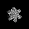 27842MC M: map data used to model this data C: citing same article ( |
|---|---|
| Similar structure data |
-Links
 PDBj
PDBj


- Assembly
| Deposited unit | 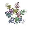
|
|---|---|
| 1 |
|
-Components
| #1: Protein | Mass: 61298.160 Da / Num. of mol.: 6 / Mutation: E325Q Source method: isolated from a genetically manipulated source Source: (gene. exp.) Lates calcarifer (barramundi perch)Production host:  References: UniProt: A0A4W6C5C5 #2: DNA chain | | Mass: 4517.935 Da / Num. of mol.: 1 / Source method: obtained synthetically / Source: (synth.) synthetic construct (others) #3: Chemical | ChemComp-ATP /   Mass: 507.181 Da / Num. of mol.: 5 / Source method: obtained synthetically / Formula: C10H16N5O13P3 / Feature type: SUBJECT OF INVESTIGATION / Comment: ATP, energy-carrying molecule*YM Mass: 507.181 Da / Num. of mol.: 5 / Source method: obtained synthetically / Formula: C10H16N5O13P3 / Feature type: SUBJECT OF INVESTIGATION / Comment: ATP, energy-carrying molecule*YM#4: Chemical | ChemComp-MG /   Mass: 24.305 Da / Num. of mol.: 5 / Source method: obtained synthetically / Formula: Mg / Feature type: SUBJECT OF INVESTIGATION Mass: 24.305 Da / Num. of mol.: 5 / Source method: obtained synthetically / Formula: Mg / Feature type: SUBJECT OF INVESTIGATIONHas ligand of interest | Y | Has protein modification | N | |
|---|
-Experimental details
-Experiment
| Experiment | Method: ELECTRON MICROSCOPY |
|---|---|
| EM experiment | Aggregation state: PARTICLE / 3D reconstruction method: single particle reconstruction |
- Sample preparation
| Component | Name: Twinkle protein, mitochondrial + DNA / Type: COMPLEX / Entity ID: #1-#2 / Source: MULTIPLE SOURCES |
|---|---|
| Source (natural) | Organism: Lates calcarifer (barramundi perch) |
| Source (recombinant) | Organism: |
| Buffer solution | pH: 8 |
| Specimen | Conc.: 0.5 mg/ml / Embedding applied: NO / Shadowing applied: NO / Staining applied: NO / Vitrification applied: YES Details: 50 mM Tris (pH 8.0), 150 mM KCl, 3 mM DTT, 1 mM ATP, and 2 mM MgCl2 |
| Specimen support | Grid material: COPPER / Grid mesh size: 300 divisions/in. / Grid type: Quantifoil R1.2/1.3 |
| Vitrification | Instrument: FEI VITROBOT MARK I / Cryogen name: ETHANE / Humidity: 100 % / Chamber temperature: 298 K |
- Electron microscopy imaging
| Experimental equipment |  Model: Titan Krios / Image courtesy: FEI Company |
|---|---|
| Microscopy | Model: FEI TITAN KRIOS |
| Electron gun | Electron source:  FIELD EMISSION GUN / Accelerating voltage: 300 kV / Illumination mode: FLOOD BEAM FIELD EMISSION GUN / Accelerating voltage: 300 kV / Illumination mode: FLOOD BEAM |
| Electron lens | Mode: BRIGHT FIELD / Nominal defocus max: 3000 nm / Nominal defocus min: 600 nm |
| Image recording | Electron dose: 49 e/Å2 / Film or detector model: GATAN K2 SUMMIT (4k x 4k) |
- Processing
| Software | Name: PHENIX / Version: 1.19.1_4122: / Classification: refinement | ||||||||||||||||||||||||
|---|---|---|---|---|---|---|---|---|---|---|---|---|---|---|---|---|---|---|---|---|---|---|---|---|---|
| EM software |
| ||||||||||||||||||||||||
| CTF correction | Type: NONE | ||||||||||||||||||||||||
| Symmetry | Point symmetry: C1 (asymmetric) | ||||||||||||||||||||||||
| 3D reconstruction | Resolution: 3.51 Å / Resolution method: FSC 0.143 CUT-OFF / Num. of particles: 44814 / Symmetry type: POINT | ||||||||||||||||||||||||
| Atomic model building | Protocol: AB INITIO MODEL / Space: REAL | ||||||||||||||||||||||||
| Refine LS restraints |
|
