 Movie
Movie Controller


+ Open data

- Basic information
| Entry | 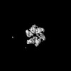 | |||||||||
|---|---|---|---|---|---|---|---|---|---|---|
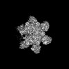
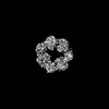
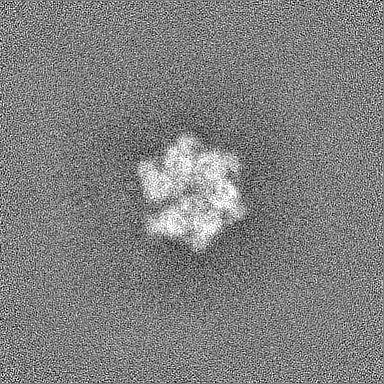
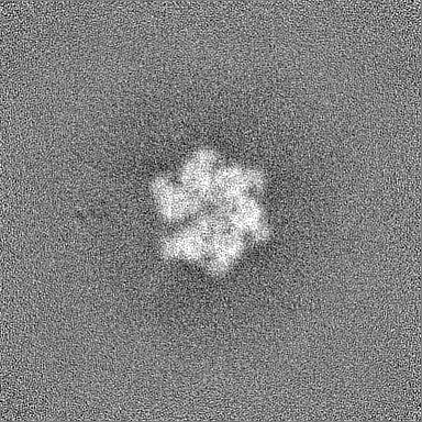
| Title | Structure of Lates calcarifer Twinkle helicase, apo hexamer | |||||||||
 Map data Map data | ||||||||||
 Sample Sample |
| |||||||||
| Biological species |  Lates calcarifer (barramundi perch) Lates calcarifer (barramundi perch) | |||||||||
| Method | single particle reconstruction / cryo EM / Resolution: 7.5 Å | |||||||||
 Authors Authors | Gao Y / Li Z | |||||||||
| Funding support |  United States, 2 items United States, 2 items
| |||||||||
 Citation Citation | Journal: Nucleic Acids Res / Year: 2022 Title: Structural and dynamic basis of DNA capture and translocation by mitochondrial Twinkle helicase. Authors: Zhuo Li / Parminder Kaur / Chen-Yu Lo / Neil Chopra / Jamie Smith / Hong Wang / Yang Gao / Abstract: Twinkle is a mitochondrial replicative helicase which can self-load onto and unwind mitochondrial DNA. Nearly 60 mutations on Twinkle have been linked to human mitochondrial diseases. Using cryo- ...Twinkle is a mitochondrial replicative helicase which can self-load onto and unwind mitochondrial DNA. Nearly 60 mutations on Twinkle have been linked to human mitochondrial diseases. Using cryo-electron microscopy (cryo-EM) and high-speed atomic force microscopy (HS-AFM), we obtained the atomic-resolution structure of a vertebrate Twinkle homolog with DNA and captured in real-time how Twinkle is self-loaded onto DNA. Our data highlight the important role of the non-catalytic N-terminal domain of Twinkle. The N-terminal domain directly contacts the C-terminal helicase domain, and the contact interface is a hotspot for disease-related mutations. Mutations at the interface destabilize Twinkle hexamer and reduce helicase activity. With HS-AFM, we observed that a highly dynamic Twinkle domain, which is likely to be the N-terminal domain, can protrude ∼5 nm to transiently capture nearby DNA and initialize Twinkle loading onto DNA. Moreover, structural analysis and subunit doping experiments suggest that Twinkle hydrolyzes ATP stochastically, which is distinct from related helicases from bacteriophages. | |||||||||
| History |
|
- Structure visualization
| Supplemental images |
|---|

- Downloads & links
-EMDB archive
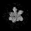
| Map data | emd_27844.map.gz | 106.4 MB |  EMDB map data format EMDB map data format | |
|---|---|---|---|---|
| Header (meta data) | emd-27844-v30.xmlemd-27844.xml | 14.7 KB 14.7 KB | Display Display | EMDB header |
| Images |  emd_27844.png emd_27844.png | 106.7 KB | ||
| Others | emd_27844_half_map_1.map.gzemd_27844_half_map_2.map.gz | 200.7 MB 200.7 MB | ||
| Archive directory |  http://ftp.pdbj.org/pub/emdb/structures/EMD-27844ftp://ftp.pdbj.org/pub/emdb/structures/EMD-27844 http://ftp.pdbj.org/pub/emdb/structures/EMD-27844ftp://ftp.pdbj.org/pub/emdb/structures/EMD-27844 | HTTPS FTP |
-Related structure data

-Links
| EMDB pages | EMDB (EBI/PDBe) / EMDataResource |
|---|
-Map
| File | Download / File: emd_27844.map.gz / Format: CCP4 / Size: 216 MB / Type: IMAGE STORED AS FLOATING POINT NUMBER (4 BYTES) | ||||||||||||||||||||||||||||||||||||
|---|---|---|---|---|---|---|---|---|---|---|---|---|---|---|---|---|---|---|---|---|---|---|---|---|---|---|---|---|---|---|---|---|---|---|---|---|---|
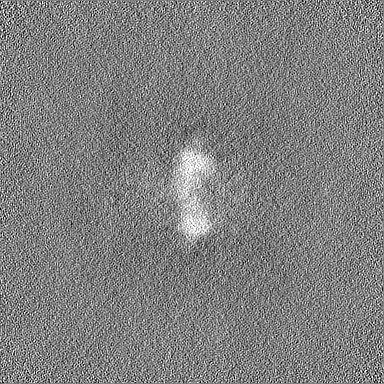
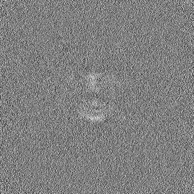
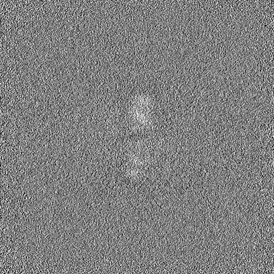
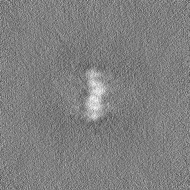
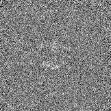
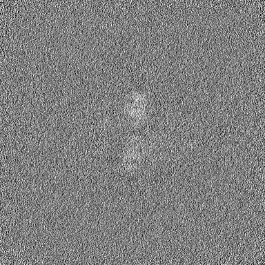
| Projections & slices | Image control
Images are generated by Spider. | ||||||||||||||||||||||||||||||||||||
| Voxel size | X=Y=Z: 1.07 Å | ||||||||||||||||||||||||||||||||||||
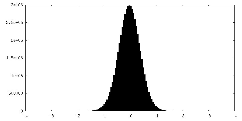
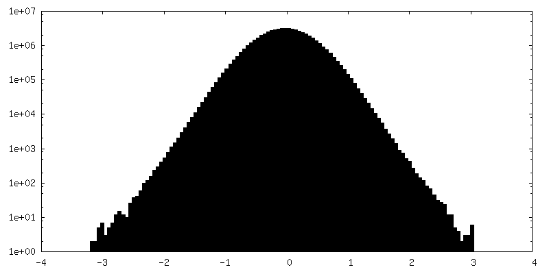
| Density |
| ||||||||||||||||||||||||||||||||||||
| Symmetry | Space group: 1 | ||||||||||||||||||||||||||||||||||||
| Details | EMDB XML:
|
 Z (Sec.)
 Y (Row.)
 X (Col.)


-Supplemental data
-Half map: #1
| File | emd_27844_half_map_1.map | ||||||||||||
|---|---|---|---|---|---|---|---|---|---|---|---|---|---|
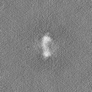
| Projections & Slices |
| ||||||||||||
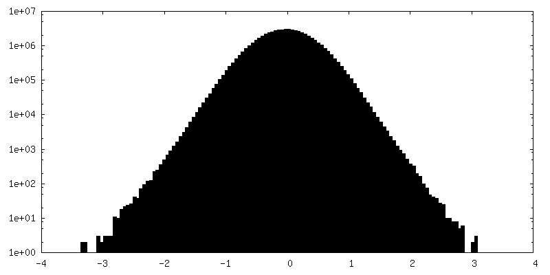
| Density Histograms |


-Half map: #2
| File | emd_27844_half_map_2.map | ||||||||||||
|---|---|---|---|---|---|---|---|---|---|---|---|---|---|
| Projections & Slices |
| ||||||||||||
| Density Histograms |


- Sample components
-Entire : Twinkle protein, mitochondrial
| Entire | Name: Twinkle protein, mitochondrial |
|---|---|
| Components |
|
-Supramolecule #1: Twinkle protein, mitochondrial
| Supramolecule | Name: Twinkle protein, mitochondrial / type: complex / ID: 1 / Chimera: Yes / Parent: 0 / Macromolecule list: all |
|---|---|
| Source (natural) | Organism: Lates calcarifer (barramundi perch) |
-Macromolecule #1: Twinkle protein, mitochondrial
| Macromolecule | Name: Twinkle protein, mitochondrial / type: protein_or_peptide / ID: 1 / Enantiomer: LEVO |
|---|---|
| Source (natural) | Organism: Lates calcarifer (barramundi perch) |
| Sequence | String: MQKEEQDFLS PHVMLGYPES LDEQEEGERE LREVQRIWSS AVPFNDLPED EAQLIKTMFQ ITKVSNATLK KFGVRLFKP TKSLVFPWFA GPDSSLKGLK LLSAQNTDTE KVTYNEATVP KISSYYNLFG LTLVGRMDSE V VLTGHELD TLAVSQATGL PSVALPRGVS ...String: MQKEEQDFLS PHVMLGYPES LDEQEEGERE LREVQRIWSS AVPFNDLPED EAQLIKTMFQ ITKVSNATLK KFGVRLFKP TKSLVFPWFA GPDSSLKGLK LLSAQNTDTE KVTYNEATVP KISSYYNLFG LTLVGRMDSE V VLTGHELD TLAVSQATGL PSVALPRGVS CLPPMLLPYL EQFKRVTLWL GHDIRSWEAS KIFSRKLGLR RC SLVRPGE DRPCPLEALA RGKNLSRIIK TSIPAAHKSI VSFKQLREDV YGELLNTEQV AGVKWTRFPE LNR ILKGHR KGELTVFTGP TGSGKTTFIS EVALDLCIQG VNTLWGSFEI NNVRLAKIML TQFAMQRLEE NLEQ YDFWA DKFEELPLYF MTFHGQQNIK TVLDTMQHAV YLYDINHVII DNLQFMMGQE NLSIDKYAVQ DHIIG AFRK FATNTSCHVT LIIHPRKEED DRELQTASIF GSAKASQEAD NVLILQEKKL VTCPGRRSLQ VTKNRF DGD VGIFPLDFIK SSLTFSAPIK GKVKLRKVST KPENEEVGGE RGGSEEGRG |
-Experimental details
-Structure determination
| Method | cryo EM |
|---|---|
 Processing Processing | single particle reconstruction |
| Aggregation state | particle |
-Sample preparation
| Concentration | 0.5 mg/mL |
|---|---|
| Buffer | pH: 8 |
| Vitrification | Cryogen name: ETHANE / Chamber humidity: 100 % / Chamber temperature: 298 K / Instrument: FEI VITROBOT MARK I |
| Details | 50 mM Tris (pH 8.0), 150 mM KCl, 3 mM DTT, 1 mM ATP, and 2 mM MgCl2 |
- Electron microscopy
| Microscope | FEI TITAN KRIOS |
|---|---|
| Image recording | Film or detector model: GATAN K2 SUMMIT (4k x 4k) / Average electron dose: 49.0 e/Å2 |
| Electron beam | Acceleration voltage: 300 kV / Electron source:  FIELD EMISSION GUN FIELD EMISSION GUN |
| Electron optics | Illumination mode: FLOOD BEAM / Imaging mode: BRIGHT FIELD / Nominal defocus max: 3.0 µm / Nominal defocus min: 0.6 µm |
| Experimental equipment |  Model: Titan Krios / Image courtesy: FEI Company |
-Image processing
| Final reconstruction | Resolution.type: BY AUTHOR / Resolution: 7.5 Å / Resolution method: FSC 0.143 CUT-OFF / Software - Name: cryoSPARC / Number images used: 10722 |
|---|---|
| Initial angle assignment | Type: MAXIMUM LIKELIHOOD / Software - Name: cryoSPARC |
| Final angle assignment | Type: MAXIMUM LIKELIHOOD / Software - Name: cryoSPARC |
-Atomic model buiding 1
| Refinement | Space: REAL / Protocol: AB INITIO MODEL |
|---|
