 Movie
Movie Controller


+ Open data

- Basic information
| Entry | Database: PDB / ID: 8.0E+19 | ||||||
|---|---|---|---|---|---|---|---|
| Title | Crystal structure of TnmK1 complexed with TNM H | ||||||
 Components Components | Secreted hydrolase | ||||||
 Keywords Keywords | HYDROLASE / biosynthesis | ||||||
| Function / homology | : / alpha/beta hydrolase fold / Alpha/beta hydrolase fold-1 / Alpha/Beta hydrolase fold / hydrolase activity / SUCCINIC ACID / Chem-U8L / Secreted hydrolase Function and homology information Function and homology information | ||||||
| Biological species |  Streptomyces sp. CB03234 (bacteria) Streptomyces sp. CB03234 (bacteria) | ||||||
| Method |  X-RAY DIFFRACTION / MOLECULAR REPLACEMENT / Resolution: 2.03 Å X-RAY DIFFRACTION / MOLECULAR REPLACEMENT / Resolution: 2.03 Å | ||||||
 Authors Authors | Liu, Y.-C. / Gui, C. / Shen, B. | ||||||
| Funding support |  United States, 1items United States, 1items
| ||||||
 Citation Citation | Journal: J.Am.Chem.Soc. / Year: 2022 Title: Intramolecular C-C Bond Formation Links Anthraquinone and Enediyne Scaffolds in Tiancimycin Biosynthesis. Authors: Gui, C. / Kalkreuter, E. / Liu, Y.C. / Adhikari, A. / Teijaro, C.N. / Yang, D. / Chang, C. / Shen, B. | ||||||
| History |
|
- Structure visualization
| Structure viewer | Molecule: MolmilJmol/JSmol |
|---|
- Downloads & links
-Download
| PDBx/mmCIF format | 8e19.cif.gz | 137.9 KB | Display | PDBx/mmCIF format |
|---|---|---|---|---|
| PDB format | pdb8e19.ent.gz | 84.8 KB | Display | PDB format |
| PDBx/mmJSON format | 8e19.json.gz | Tree view | PDBx/mmJSON format | |
| Others |  Other downloads Other downloads |
-Validation report
| Summary document | 8e19_validation.pdf.gz | 844.1 KB | Display | wwPDB validaton report |
|---|---|---|---|---|
| Full document | 8e19_full_validation.pdf.gz | 849 KB | Display | |
| Data in XML | 8e19_validation.xml.gz | 22.8 KB | Display | |
| Data in CIF | 8e19_validation.cif.gz | 33.9 KB | Display | |
| Arichive directory | https://data.pdbj.org/pub/pdb/validation_reports/e1/8e19ftp://data.pdbj.org/pub/pdb/validation_reports/e1/8e19 | HTTPS FTP |
-Related structure data
| Related structure data |  8e18SC S: Starting model for refinement C: citing same article ( |
|---|---|
| Similar structure data |
-Links
 PDBj
PDBj


- Assembly
| Deposited unit | 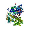
| ||||||||||||
|---|---|---|---|---|---|---|---|---|---|---|---|---|---|
| 1 |
| ||||||||||||
| Unit cell |
| ||||||||||||
| Components on special symmetry positions |
|
-Components
| #1: Protein | Mass: 52201.891 Da / Num. of mol.: 1 Source method: isolated from a genetically manipulated source Source: (gene. exp.) Streptomyces sp. CB03234 (bacteria) / Gene: tnmK1, AMK26_32035 / Production host: Streptomyces lividans (bacteria) / References: UniProt: A0A125SA14 |
|---|---|
| #2: Chemical | ChemComp-U8L / (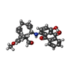  Mass: 477.507 Da / Num. of mol.: 1 / Source method: obtained synthetically / Formula: C30H23NO5 / Feature type: SUBJECT OF INVESTIGATION Mass: 477.507 Da / Num. of mol.: 1 / Source method: obtained synthetically / Formula: C30H23NO5 / Feature type: SUBJECT OF INVESTIGATION |
| #3: Chemical | ChemComp-EPE /   Mass: 238.305 Da / Num. of mol.: 1 / Source method: obtained synthetically / Formula: C8H18N2O4S / Comment: pH buffer*YM Mass: 238.305 Da / Num. of mol.: 1 / Source method: obtained synthetically / Formula: C8H18N2O4S / Comment: pH buffer*YM |
| #4: Chemical | ChemComp-SIN /   Mass: 118.088 Da / Num. of mol.: 1 / Source method: obtained synthetically / Formula: C4H6O4 Mass: 118.088 Da / Num. of mol.: 1 / Source method: obtained synthetically / Formula: C4H6O4 |
| #5: Water | ChemComp-HOH /  Mass: 18.015 Da / Num. of mol.: 377 / Source method: isolated from a natural source / Formula: H2O Mass: 18.015 Da / Num. of mol.: 377 / Source method: isolated from a natural source / Formula: H2O |
| Has ligand of interest | Y |
-Experimental details
-Experiment
| Experiment | Method: X-RAY DIFFRACTION / Number of used crystals: 1 |
|---|
- Sample preparation
| Crystal | Density Matthews: 2.28 Å3/Da / Density % sol: 46.1 % |
|---|---|
| Crystal grow | Temperature: 298 K / Method: vapor diffusion, sitting drop / pH: 7 Details: 0.1 M HEPES, pH 7.0, 1.0 M succinic acid, pH 7.0, and 1% w/v PEG 2000 MME |
-Data collection
| Diffraction | Mean temperature: 100 K / Serial crystal experiment: N |
|---|---|
| Diffraction source | Source: ROTATING ANODE / Type: RIGAKU MICROMAX-007 HF / Wavelength: 1.54 Å |
| Detector | Type: MAR scanner 345 mm plate / Detector: IMAGE PLATE / Date: Dec 10, 2019 |
| Radiation | Protocol: SINGLE WAVELENGTH / Monochromatic (M) / Laue (L): M / Scattering type: x-ray |
| Radiation wavelength | Wavelength: 1.54 Å / Relative weight: 1 |
| Reflection | Resolution: 2.03→30 Å / Num. obs: 31893 / % possible obs: 100 % / Redundancy: 13.2 % / Biso Wilson estimate: 27.12 Å2 / CC1/2: 0.998 / Net I/σ(I): 32 |
| Reflection shell | Resolution: 2.03→2.06 Å / Num. unique obs: 1569 / CC1/2: 0.728 |
- Processing
| Software |
| ||||||||||||||||||||||||||||||||||||||||||||||||||||||||||||||||||||||||||||||||||||
|---|---|---|---|---|---|---|---|---|---|---|---|---|---|---|---|---|---|---|---|---|---|---|---|---|---|---|---|---|---|---|---|---|---|---|---|---|---|---|---|---|---|---|---|---|---|---|---|---|---|---|---|---|---|---|---|---|---|---|---|---|---|---|---|---|---|---|---|---|---|---|---|---|---|---|---|---|---|---|---|---|---|---|---|---|---|
| Refinement | Method to determine structure: MOLECULAR REPLACEMENT / Starting model: 8.0E+18 / Resolution: 2.03→29.55 Å / SU ML: 0.2103 / Cross valid method: FREE R-VALUE / σ(F): 1.33 / Phase error: 19.6859 Stereochemistry target values: GeoStd + Monomer Library + CDL v1.2
| ||||||||||||||||||||||||||||||||||||||||||||||||||||||||||||||||||||||||||||||||||||
| Solvent computation | Shrinkage radii: 0.9 Å / VDW probe radii: 1.11 Å / Solvent model: FLAT BULK SOLVENT MODEL | ||||||||||||||||||||||||||||||||||||||||||||||||||||||||||||||||||||||||||||||||||||
| Displacement parameters | Biso mean: 30.5 Å2 | ||||||||||||||||||||||||||||||||||||||||||||||||||||||||||||||||||||||||||||||||||||
| Refinement step | Cycle: LAST / Resolution: 2.03→29.55 Å
| ||||||||||||||||||||||||||||||||||||||||||||||||||||||||||||||||||||||||||||||||||||
| Refine LS restraints |
| ||||||||||||||||||||||||||||||||||||||||||||||||||||||||||||||||||||||||||||||||||||
| LS refinement shell |
|
