 ムービー
ムービー コントローラー


+ データを開く

- 基本情報
基本情報
| 登録情報 | データベース: PDB / ID: 8.0E+15 | |||||||||
|---|---|---|---|---|---|---|---|---|---|---|
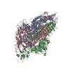
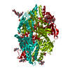
| タイトル | A computationally stabilized hMPV F protein | |||||||||
 要素 要素 |
| |||||||||
 キーワード キーワード | VIRAL PROTEIN / Human metapneumovirus / fusion protein / computational stabilization | |||||||||
| 機能・相同性 |  機能・相同性情報 機能・相同性情報fusion of virus membrane with host plasma membrane / viral envelope / symbiont entry into host cell / host cell plasma membrane / virion membrane / membrane 類似検索 - 分子機能 | |||||||||
| 生物種 |  Human metapneumovirus (ウイルス)Enterobacteria phage T2 (ファージ) Human metapneumovirus (ウイルス)Enterobacteria phage T2 (ファージ) | |||||||||
| 手法 |  X線回折 / シンクロトロン / 分子置換 / 解像度: 2.41 Å X線回折 / シンクロトロン / 分子置換 / 解像度: 2.41 Å | |||||||||
 データ登録者 データ登録者 | Huang, J. / Gonzalez, K. / Mousa, J. / Strauch, E. | |||||||||
| 資金援助 |  米国, 2件 米国, 2件
| |||||||||
 引用 引用 | ジャーナル: Nat Commun / 年: 2024 タイトル: A general computational design strategy for stabilizing viral class I fusion proteins. 著者: Karen J Gonzalez / Jiachen Huang / Miria F Criado / Avik Banerjee / Stephen M Tompkins / Jarrod J Mousa / Eva-Maria Strauch / 要旨: Many pathogenic viruses rely on class I fusion proteins to fuse their viral membrane with the host cell membrane. To drive the fusion process, class I fusion proteins undergo an irreversible ...Many pathogenic viruses rely on class I fusion proteins to fuse their viral membrane with the host cell membrane. To drive the fusion process, class I fusion proteins undergo an irreversible conformational change from a metastable prefusion state to an energetically more stable postfusion state. Mounting evidence underscores that antibodies targeting the prefusion conformation are the most potent, making it a compelling vaccine candidate. Here, we establish a computational design protocol that stabilizes the prefusion state while destabilizing the postfusion conformation. With this protocol, we stabilize the fusion proteins of the RSV, hMPV, and SARS-CoV-2 viruses, testing fewer than a handful of designs. The solved structures of these designed proteins from all three viruses evidence the atomic accuracy of our approach. Furthermore, the humoral response of the redesigned RSV F protein compares to that of the recently approved vaccine in a mouse model. While the parallel design of two conformations allows the identification of energetically sub-optimal positions for one conformation, our protocol also reveals diverse molecular strategies for stabilization. Given the clinical significance of viruses using class I fusion proteins, our algorithm can substantially contribute to vaccine development by reducing the time and resources needed to optimize these immunogens. #1: ジャーナル: bioRxiv / 年: 2023 タイトル: A general computational design strategy for stabilizing viral class I fusion proteins. 著者: Karen J Gonzalez / Jiachen Huang / Miria F Criado / Avik Banerjee / Stephen Tompkins / Jarrod J Mousa / Eva-Maria Strauch / 要旨: Many pathogenic viruses, including influenza virus, Ebola virus, coronaviruses, and Pneumoviruses, rely on class I fusion proteins to fuse viral and cellular membranes. To drive the fusion process, ...Many pathogenic viruses, including influenza virus, Ebola virus, coronaviruses, and Pneumoviruses, rely on class I fusion proteins to fuse viral and cellular membranes. To drive the fusion process, class I fusion proteins undergo an irreversible conformational change from a metastable prefusion state to an energetically more favorable and stable postfusion state. An increasing amount of evidence exists highlighting that antibodies targeting the prefusion conformation are the most potent. However, many mutations have to be evaluated before identifying prefusion-stabilizing substitutions. We therefore established a computational design protocol that stabilizes the prefusion state while destabilizing the postfusion conformation. As a proof of concept, we applied this principle to the fusion protein of the RSV, hMPV, and SARS-CoV-2 viruses. For each protein, we tested less than a handful of designs to identify stable versions. Solved structures of designed proteins from the three different viruses evidenced the atomic accuracy of our approach. Furthermore, the immunological response of the RSV F design compared to a current clinical candidate in a mouse model. While the parallel design of two conformations allows identifying and selectively modifying energetically less optimized positions for one conformation, our protocol also reveals diverse molecular strategies for stabilization. We recaptured many approaches previously introduced manually for the stabilization of viral surface proteins, such as cavity-filling, optimization of polar interactions, as well as postfusion-disruptive strategies. Using our approach, it is possible to focus on the most impacting mutations and potentially preserve the immunogen as closely as possible to its native version. The latter is important as sequence re-design can cause perturbations to B and T cell epitopes. Given the clinical significance of viruses using class I fusion proteins, our algorithm can substantially contribute to vaccine development by reducing the time and resources needed to optimize these immunogens. | |||||||||
| 履歴 |
|
- 構造の表示
構造の表示
| 構造ビューア | 分子: MolmilJmol/JSmol |
|---|
- ダウンロードとリンク
-ダウンロード
| PDBx/mmCIF形式 | 8e15.cif.gz | 105.8 KB | 表示 | PDBx/mmCIF形式 |
|---|---|---|---|---|
| PDB形式 | pdb8e15.ent.gz | 75.9 KB | 表示 | PDB形式 |
| PDBx/mmJSON形式 | 8e15.json.gz | ツリー表示 | PDBx/mmJSON形式 | |
| その他 |  その他のダウンロード その他のダウンロード |
-検証レポート
| 文書・要旨 | 8e15_validation.pdf.gz | 788.9 KB | 表示 | wwPDB検証レポート |
|---|---|---|---|---|
| 文書・詳細版 | 8e15_full_validation.pdf.gz | 792.5 KB | 表示 | |
| XML形式データ | 8e15_validation.xml.gz | 17.6 KB | 表示 | |
| CIF形式データ | 8e15_validation.cif.gz | 23.5 KB | 表示 | |
| アーカイブディレクトリ | https://data.pdbj.org/pub/pdb/validation_reports/e1/8e15ftp://data.pdbj.org/pub/pdb/validation_reports/e1/8e15 | HTTPS FTP |
-関連構造データ


-リンク
 PDBj
PDBj


- 集合体
集合体
| 登録構造単位 | 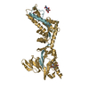
| ||||||||||||
|---|---|---|---|---|---|---|---|---|---|---|---|---|---|
| 1 | 
| ||||||||||||
| 単位格子 |
|
-要素
| #1: タンパク質 | 分子量: 11687.245 Da / 分子数: 1 / 由来タイプ: 組換発現 / 由来: (組換発現) Human metapneumovirus (ウイルス) / 細胞株 (発現宿主): 293 / 発現宿主:  Homo sapiens (ヒト) / 参照: UniProt: Q8B9P0 Homo sapiens (ヒト) / 参照: UniProt: Q8B9P0 | ||||||
|---|---|---|---|---|---|---|---|
| #2: タンパク質 | 分子量: 45999.160 Da / 分子数: 1 / 由来タイプ: 組換発現 由来: (組換発現) Human metapneumovirus (ウイルス), (組換発現) Enterobacteria phage T2 (ファージ)遺伝子: F, wac, EcT2_00172 / 細胞株 (発現宿主): 293 / 発現宿主: Homo sapiens (ヒト) / 参照: UniProt: Q8B9P0, UniProt: Q76VI8 | ||||||
| #3: 多糖 | beta-D-mannopyranose-(1-4)-2-acetamido-2-deoxy-beta-D-glucopyranose-(1-4)-2-acetamido-2-deoxy-beta- ...beta-D-mannopyranose-(1-4)-2-acetamido-2-deoxy-beta-D-glucopyranose-(1-4)-2-acetamido-2-deoxy-beta-D-glucopyranose | ||||||
| #4: 糖 |   タイプ: D-saccharide, beta linking / 分子量: 221.208 Da / 分子数: 2 / 由来タイプ: 合成 / 式: C8H15NO6 タイプ: D-saccharide, beta linking / 分子量: 221.208 Da / 分子数: 2 / 由来タイプ: 合成 / 式: C8H15NO6#5: 水 | ChemComp-HOH / |  分子量: 18.015 Da / 分子数: 11 / 由来タイプ: 天然 / 式: H2O 分子量: 18.015 Da / 分子数: 11 / 由来タイプ: 天然 / 式: H2O研究の焦点であるリガンドがあるか | N | Has protein modification | Y | |
-実験情報
-実験
| 実験 | 手法: X線回折 / 使用した結晶の数: 1 |
|---|
- 試料調製
試料調製
| 結晶 | マシュー密度: 4.09 Å3/Da / 溶媒含有率: 69.91 % |
|---|---|
| 結晶化 | 温度: 298 K / 手法: 蒸気拡散法, シッティングドロップ法 詳細: 0.1 M Sodium acetate trihydrate pH 4.6, 2.0 M Sodium formate |
-データ収集
| 回折 | 平均測定温度: 100 K / Serial crystal experiment: N |
|---|---|
| 放射光源 | 由来: シンクロトロン / サイト: APS / ビームライン: 21-ID-D / 波長: 1 Å |
| 検出器 | タイプ: DECTRIS EIGER X 16M / 検出器: PIXEL / 日付: 2022年4月21日 |
| 放射 | プロトコル: SINGLE WAVELENGTH / 単色(M)・ラウエ(L): M / 散乱光タイプ: x-ray |
| 放射波長 | 波長: 1 Å / 相対比: 1 |
| 反射 | 解像度: 2.41→47.62 Å / Num. obs: 36363 / % possible obs: 99.91 % / 冗長度: 2 % / Biso Wilson estimate: 66.92 Å2 / CC1/2: 0.999 / CC star: 1 / Net I/σ(I): 9.18 |
| 反射 シェル | 解像度: 2.41→2.496 Å / 冗長度: 2 % / Mean I/σ(I) obs: 0.78 / Num. unique obs: 3606 / CC1/2: 0.593 / CC star: 0.863 / % possible all: 99.86 |
- 解析
解析
| ソフトウェア |
| ||||||||||||||||||||||||||||||||||||||||||||||||||||||||||||||||||||||||||||||||||||||||||||||||||
|---|---|---|---|---|---|---|---|---|---|---|---|---|---|---|---|---|---|---|---|---|---|---|---|---|---|---|---|---|---|---|---|---|---|---|---|---|---|---|---|---|---|---|---|---|---|---|---|---|---|---|---|---|---|---|---|---|---|---|---|---|---|---|---|---|---|---|---|---|---|---|---|---|---|---|---|---|---|---|---|---|---|---|---|---|---|---|---|---|---|---|---|---|---|---|---|---|---|---|---|
| 精密化 | 構造決定の手法: 分子置換 開始モデル: 5wb0 解像度: 2.41→47.62 Å / SU ML: 0.3364 / 交差検証法: FREE R-VALUE / σ(F): 1.34 / 位相誤差: 27.647 立体化学のターゲット値: GeoStd + Monomer Library + CDL v1.2
| ||||||||||||||||||||||||||||||||||||||||||||||||||||||||||||||||||||||||||||||||||||||||||||||||||
| 溶媒の処理 | 減衰半径: 0.9 Å / VDWプローブ半径: 1.1 Å / 溶媒モデル: FLAT BULK SOLVENT MODEL | ||||||||||||||||||||||||||||||||||||||||||||||||||||||||||||||||||||||||||||||||||||||||||||||||||
| 原子変位パラメータ | Biso mean: 70.9 Å2 | ||||||||||||||||||||||||||||||||||||||||||||||||||||||||||||||||||||||||||||||||||||||||||||||||||
| 精密化ステップ | サイクル: LAST / 解像度: 2.41→47.62 Å
| ||||||||||||||||||||||||||||||||||||||||||||||||||||||||||||||||||||||||||||||||||||||||||||||||||
| 拘束条件 |
| ||||||||||||||||||||||||||||||||||||||||||||||||||||||||||||||||||||||||||||||||||||||||||||||||||
| LS精密化 シェル |
|
