- PDB-8e11: Structure of mouse DNA polymerase Beta (PolB) mutant -
+
Open data
ID or keywords:
Loading...
-
Basic information
Entry
Database: PDB / ID: 800000000000
Title
Structure of mouse DNA polymerase Beta (PolB) mutant
Components
DNA polymerase beta
Keywords
TRANSFERASE / DNA Polymerase / Base Excision Repair / DNA binding
Function / homology
Function and homology information
Resolution of AP sites via the multiple-nucleotide patch replacement pathway / Abasic sugar-phosphate removal via the single-nucleotide replacement pathway / Resolution of AP sites via the single-nucleotide replacement pathway / PCNA-Dependent Long Patch Base Excision Repair / POLB-Dependent Long Patch Base Excision Repair / APEX1-Independent Resolution of AP Sites via the Single Nucleotide Replacement Pathway / somatic diversification of immunoglobulins / synaptonemal complex / Ub-specific processing proteases / immunoglobulin heavy chain V-D-J recombination ...Resolution of AP sites via the multiple-nucleotide patch replacement pathway / Abasic sugar-phosphate removal via the single-nucleotide replacement pathway / Resolution of AP sites via the single-nucleotide replacement pathway / PCNA-Dependent Long Patch Base Excision Repair / POLB-Dependent Long Patch Base Excision Repair / APEX1-Independent Resolution of AP Sites via the Single Nucleotide Replacement Pathway / somatic diversification of immunoglobulins / synaptonemal complex / Ub-specific processing proteases / immunoglobulin heavy chain V-D-J recombination / Lyases; Carbon-oxygen lyases; Other carbon-oxygen lyases / pyrimidine dimer repair / homeostasis of number of cells / response to hyperoxia / 5'-deoxyribose-5-phosphate lyase activity / lymph node development / salivary gland morphogenesis / somatic hypermutation of immunoglobulin genes / spleen development / base-excision repair, gap-filling / class I DNA-(apurinic or apyrimidinic site) endonuclease activity / DNA-(apurinic or apyrimidinic site) lyase / response to gamma radiation / spindle microtubule / base-excision repair / intrinsic apoptotic signaling pathway in response to DNA damage / neuron apoptotic process / microtubule binding / DNA-directed DNA polymerase / response to ethanol / in utero embryonic development / microtubule / damaged DNA binding / DNA-directed DNA polymerase activity / DNA replication / lyase activity / inflammatory response / apoptotic process / DNA damage response / enzyme binding / protein-containing complex / metal ion binding / cytoplasm Similarity search - Function
Beta Polymerase; domain 3 / DNA polymerase, thumb domain / DNA polymerase family X, beta-like / DNA polymerase beta, palm domain / DNA polymerase beta palm / DNA polymerase lambda, fingers domain / Fingers domain of DNA polymerase lambda / DNA-directed DNA polymerase X / DNA polymerase X family / DNA polymerase beta-like, N-terminal domain ...Beta Polymerase; domain 3 / DNA polymerase, thumb domain / DNA polymerase family X, beta-like / DNA polymerase beta, palm domain / DNA polymerase beta palm / DNA polymerase lambda, fingers domain / Fingers domain of DNA polymerase lambda / DNA-directed DNA polymerase X / DNA polymerase X family / DNA polymerase beta-like, N-terminal domain / Helix-hairpin-helix domain / DNA polymerase family X, binding site / DNA polymerase family X signature. / DNA polymerase lambda lyase domain superfamily / DNA polymerase family X / DNA polymerase beta, thumb domain / DNA polymerase, thumb domain superfamily / DNA polymerase beta thumb / Beta Polymerase, domain 2 / Helix-hairpin-helix DNA-binding motif, class 1 / Helix-hairpin-helix DNA-binding motif class 1 / Beta Polymerase; domain 2 / Nucleotidyltransferase superfamily / 5' to 3' exonuclease, C-terminal subdomain / DNA polymerase; domain 1 / 2-Layer Sandwich / Orthogonal Bundle / Mainly Alpha / Alpha Beta Similarity search - Domain/homology
 Movie
Movie Controller


 Open data
 Basic information
 Components
 Keywords
 Function and homology information
 X-RAY DIFFRACTION /
  Authors
 United States, 1items
  Citation
 Structure visualization
 Downloads & links
 Other downloads
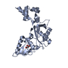

 PDBj
PDBj


 Assembly


