 Movie
Movie Controller

[English] 日本語
 Yorodumi
- PDB-8e02: Crystal structure of photoactive yellow protein (PYP); F96oCNF T1... -
+ Open data

- Basic information
| Entry | Database: PDB / ID: 800 | |||||||||
|---|---|---|---|---|---|---|---|---|---|---|
| Title | Crystal structure of photoactive yellow protein (PYP); F96oCNF T103V construct | |||||||||
 Components Components | Photoactive yellow protein | |||||||||
 Keywords Keywords | SIGNALING PROTEIN / PHOTORECEPTOR | |||||||||
| Function / homology |  Function and homology information Function and homology informationphototransduction / photoreceptor activity / regulation of DNA-templated transcription / identical protein binding Similarity search - Function | |||||||||
| Biological species |  Halorhodospira halophila (bacteria) Halorhodospira halophila (bacteria) | |||||||||
| Method |  X-RAY DIFFRACTION / SYNCHROTRON / MOLECULAR REPLACEMENT / Resolution: 1.09 Å X-RAY DIFFRACTION / SYNCHROTRON / MOLECULAR REPLACEMENT / Resolution: 1.09 Å | |||||||||
 Authors Authors | Kirsh, J.M. / Weaver, J.B. / Boxer, S.G. | |||||||||
| Funding support |  United States, 2items United States, 2items
| |||||||||
 Citation Citation | Journal: To Be Published Title: Protic and Aprotic Interactions Systematically Perturbed and Mapped via MD and IR Spectroscopy Authors: Kirsh, J.M. / Kozuch, J. / Weaver, J.B. / Boxer, S.G. | |||||||||
| History |
|
- Structure visualization
| Structure viewer | Molecule: MolmilJmol/JSmol |
|---|
- Downloads & links
-Download
| PDBx/mmCIF format | 8e02.cif.gz | 75.7 KB | Display | PDBx/mmCIF format |
|---|---|---|---|---|
| PDB format | pdb8e02.ent.gz | 53.6 KB | Display | PDB format |
| PDBx/mmJSON format | 8e02.json.gz | Tree view | PDBx/mmJSON format | |
| Others |  Other downloads Other downloads |
-Validation report
| Summary document | 8e02_validation.pdf.gz | 441.9 KB | Display | wwPDB validaton report |
|---|---|---|---|---|
| Full document | 8e02_full_validation.pdf.gz | 443.1 KB | Display | |
| Data in XML | 8e02_validation.xml.gz | 8.7 KB | Display | |
| Data in CIF | 8e02_validation.cif.gz | 11.8 KB | Display | |
| Arichive directory | https://data.pdbj.org/pub/pdb/validation_reports/e0/8e02ftp://data.pdbj.org/pub/pdb/validation_reports/e0/8e02 | HTTPS FTP |
-Related structure data
| Related structure data |  8dzuC  8dzxC  8dzyC  8e03C  8e09C  8e1kC  8e1lC 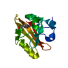 1nwzS S: Starting model for refinement C: citing same article ( |
|---|---|
| Similar structure data |
-Links
 PDBj
PDBj


- Assembly
| Deposited unit | 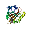
| ||||||||
|---|---|---|---|---|---|---|---|---|---|
| 1 |
| ||||||||
| Unit cell |
|
-Components
| #1: Protein | Mass: 13911.612 Da / Num. of mol.: 1 Source method: isolated from a genetically manipulated source Source: (gene. exp.) Halorhodospira halophila (bacteria) / Gene: pypProduction host: Strain (production host): DH10B / References: UniProt: P16113 |
|---|---|
| #2: Chemical | ChemComp-HC4 /   Mass: 164.158 Da / Num. of mol.: 1 / Source method: obtained synthetically / Formula: C9H8O3 Mass: 164.158 Da / Num. of mol.: 1 / Source method: obtained synthetically / Formula: C9H8O3 |
| #3: Water | ChemComp-HOH /  Mass: 18.015 Da / Num. of mol.: 121 / Source method: isolated from a natural source / Formula: H2O Mass: 18.015 Da / Num. of mol.: 121 / Source method: isolated from a natural source / Formula: H2O |
| Has ligand of interest | Y |
-Experimental details
-Experiment
| Experiment | Method: X-RAY DIFFRACTION / Number of used crystals: 1 |
|---|
- Sample preparation
| Crystal | Density Matthews: 1.84 Å3/Da / Density % sol: 33.12 % |
|---|---|
| Crystal grow | Temperature: 293 K / Method: vapor diffusion, hanging drop / pH: 6 Details: 20 mM potassium phosphate, pH 6.0, 1 M NaCl; 2.0 M ammonium sulfate |
-Data collection
| Diffraction | Mean temperature: 100 K / Serial crystal experiment: N |
|---|---|
| Diffraction source | Source: SYNCHROTRON / Site: SSRL / Beamline: BL12-1 / Wavelength: 0.7126 Å |
| Detector | Type: DECTRIS EIGER2 XE 16M / Detector: PIXEL / Date: Feb 26, 2021 |
| Radiation | Protocol: SINGLE WAVELENGTH / Monochromatic (M) / Laue (L): M / Scattering type: x-ray |
| Radiation wavelength | Wavelength: 0.7126 Å / Relative weight: 1 |
| Reflection | Resolution: 1.09→33.2 Å / Num. obs: 41825 / % possible obs: 98.5 % / Redundancy: 41.4 % / Biso Wilson estimate: 9.94 Å2 / CC1/2: 1 / Rmerge(I) obs: 0.178 / Rpim(I) all: 0.028 / Rrim(I) all: 0.18 / Net I/σ(I): 15.8 / Num. measured all: 1730378 / Scaling rejects: 79 |
| Reflection shell | Resolution: 1.09→1.11 Å / Redundancy: 24.3 % / Rmerge(I) obs: 4.225 / Num. measured all: 35988 / Num. unique obs: 1483 / CC1/2: 0.386 / Rpim(I) all: 0.855 / Rrim(I) all: 4.32 / Net I/σ(I) obs: 1.6 / % possible all: 70.7 |
- Processing
| Software |
| ||||||||||||||||||||||||||||||||||||||||||||||||||||||||||||||||||||||||||||||||||||||||||||||||||||||||||||||||
|---|---|---|---|---|---|---|---|---|---|---|---|---|---|---|---|---|---|---|---|---|---|---|---|---|---|---|---|---|---|---|---|---|---|---|---|---|---|---|---|---|---|---|---|---|---|---|---|---|---|---|---|---|---|---|---|---|---|---|---|---|---|---|---|---|---|---|---|---|---|---|---|---|---|---|---|---|---|---|---|---|---|---|---|---|---|---|---|---|---|---|---|---|---|---|---|---|---|---|---|---|---|---|---|---|---|---|---|---|---|---|---|---|---|
| Refinement | Method to determine structure: MOLECULAR REPLACEMENT Starting model: 1NWZ Resolution: 1.09→28.71 Å / SU ML: 0.09 / Cross valid method: THROUGHOUT / σ(F): 1.34 / Phase error: 17.37 / Stereochemistry target values: ML
| ||||||||||||||||||||||||||||||||||||||||||||||||||||||||||||||||||||||||||||||||||||||||||||||||||||||||||||||||
| Solvent computation | Shrinkage radii: 0.9 Å / VDW probe radii: 1.11 Å / Solvent model: FLAT BULK SOLVENT MODEL | ||||||||||||||||||||||||||||||||||||||||||||||||||||||||||||||||||||||||||||||||||||||||||||||||||||||||||||||||
| Displacement parameters | Biso max: 43.01 Å2 / Biso mean: 13.52 Å2 / Biso min: 3.73 Å2 | ||||||||||||||||||||||||||||||||||||||||||||||||||||||||||||||||||||||||||||||||||||||||||||||||||||||||||||||||
| Refinement step | Cycle: final / Resolution: 1.09→28.71 Å
| ||||||||||||||||||||||||||||||||||||||||||||||||||||||||||||||||||||||||||||||||||||||||||||||||||||||||||||||||
| LS refinement shell | Refine-ID: X-RAY DIFFRACTION / Rfactor Rfree error: 0 / Total num. of bins used: 15
|
