 Movie
Movie Controller

[English] 日本語
 Yorodumi
- PDB-8dw7: DNA glycosylase MutY variant N146S in complex with DNA containing... -
+ Open data

- Basic information
| Entry | Database: PDB / ID: 8dw7 | |||||||||||||||
|---|---|---|---|---|---|---|---|---|---|---|---|---|---|---|---|---|
| Title | DNA glycosylase MutY variant N146S in complex with DNA containing the transition state analog 1N paired with d(8-oxo-G) | |||||||||||||||
 Components Components |
| |||||||||||||||
 Keywords Keywords | HYDROLASE/DNA / Protein-DNA complex / DNA repair / transition state / Base Excision Repair / HYDROLASE / HYDROLASE-DNA complex | |||||||||||||||
| Function / homology |  Function and homology information Function and homology informationadenine/guanine mispair binding / adenine glycosylase / DNA N-glycosylase activity / 8-oxo-7,8-dihydroguanine DNA N-glycosylase activity / purine-specific mismatch base pair DNA N-glycosylase activity / oxidized purine DNA binding / mismatch repair / base-excision repair / 4 iron, 4 sulfur cluster binding / DNA binding / metal ion binding Similarity search - Function | |||||||||||||||
| Biological species |   Geobacillus stearothermophilus (bacteria) Geobacillus stearothermophilus (bacteria)synthetic construct (others) | |||||||||||||||
| Method |  X-RAY DIFFRACTION / SYNCHROTRON / MOLECULAR REPLACEMENT / Resolution: 1.96 Å X-RAY DIFFRACTION / SYNCHROTRON / MOLECULAR REPLACEMENT / Resolution: 1.96 Å | |||||||||||||||
 Authors Authors | Demir, M. / Russelburg, L.P. / Horvath, M.P. / David, S.S. | |||||||||||||||
| Funding support |  United States, 4items United States, 4items
| |||||||||||||||
 Citation Citation | Journal: Nucleic Acids Res. / Year: 2023 Title: Structural snapshots of base excision by the cancer-associated variant MutY N146S reveal a retaining mechanism. Authors: Demir, M. / Russelburg, L.P. / Lin, W.J. / Trasvina-Arenas, C.H. / Huang, B. / Yuen, P.K. / Horvath, M.P. / David, S.S. | |||||||||||||||
| History |
|
- Structure visualization
| Structure viewer | Molecule: MolmilJmol/JSmol |
|---|
- Downloads & links
-Download
| PDBx/mmCIF format | 8dw7.cif.gz | 546.2 KB | Display | PDBx/mmCIF format |
|---|---|---|---|---|
| PDB format | pdb8dw7.ent.gz | 374.4 KB | Display | PDB format |
| PDBx/mmJSON format | 8dw7.json.gz | Tree view | PDBx/mmJSON format | |
| Others |  Other downloads Other downloads |
-Validation report
| Summary document | 8dw7_validation.pdf.gz | 2.2 MB | Display | wwPDB validaton report |
|---|---|---|---|---|
| Full document | 8dw7_full_validation.pdf.gz | 2.2 MB | Display | |
| Data in XML | 8dw7_validation.xml.gz | 31.3 KB | Display | |
| Data in CIF | 8dw7_validation.cif.gz | 44 KB | Display | |
| Arichive directory | https://data.pdbj.org/pub/pdb/validation_reports/dw/8dw7ftp://data.pdbj.org/pub/pdb/validation_reports/dw/8dw7 | HTTPS FTP |
-Related structure data
| Related structure data | 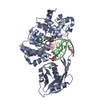 8dvpC  8dvyC 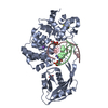 8dw0C  8dw4C 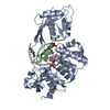 6u7tS C: citing same article ( S: Starting model for refinement |
|---|---|
| Similar structure data |
-Links
 PDBj
PDBj


- Assembly
| Deposited unit | 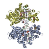
| ||||||||||||
|---|---|---|---|---|---|---|---|---|---|---|---|---|---|
| 1 | 
| ||||||||||||
| 2 | 
| ||||||||||||
| Unit cell |
|
-Components
-Protein , 1 types, 2 molecules AD
| #1: Protein | Mass: 41746.574 Da / Num. of mol.: 2 / Mutation: N146S Source method: isolated from a genetically manipulated source Source: (gene. exp.) Geobacillus stearothermophilus (bacteria)Gene: mutY / Production host: |
|---|
-DNA chain , 2 types, 4 molecules BECF
| #2: DNA chain | Mass: 3423.249 Da / Num. of mol.: 2 / Source method: obtained synthetically / Source: (synth.) synthetic construct (others) #3: DNA chain | Mass: 3191.096 Da / Num. of mol.: 2 / Source method: obtained synthetically / Source: (synth.) synthetic construct (others) |
|---|
-Non-polymers , 5 types, 198 molecules 


| #4: Chemical |  Mass: 351.640 Da / Num. of mol.: 2 / Source method: obtained synthetically / Formula: Fe4S4 / Feature type: SUBJECT OF INVESTIGATION Mass: 351.640 Da / Num. of mol.: 2 / Source method: obtained synthetically / Formula: Fe4S4 / Feature type: SUBJECT OF INVESTIGATION#5: Chemical |  Mass: 62.068 Da / Num. of mol.: 2 / Source method: obtained synthetically / Formula: C2H6O2 Mass: 62.068 Da / Num. of mol.: 2 / Source method: obtained synthetically / Formula: C2H6O2#6: Chemical |  Mass: 40.078 Da / Num. of mol.: 2 / Source method: obtained synthetically / Formula: Ca / Feature type: SUBJECT OF INVESTIGATION Mass: 40.078 Da / Num. of mol.: 2 / Source method: obtained synthetically / Formula: Ca / Feature type: SUBJECT OF INVESTIGATION#7: Chemical |  Mass: 122.143 Da / Num. of mol.: 2 / Source method: obtained synthetically / Formula: C4H12NO3 / Comment: pH buffer*YM Mass: 122.143 Da / Num. of mol.: 2 / Source method: obtained synthetically / Formula: C4H12NO3 / Comment: pH buffer*YM#8: Water | ChemComp-HOH / | Mass: 18.015 Da / Num. of mol.: 190 / Source method: isolated from a natural source / Formula: H2O |
|---|
-Details
| Has ligand of interest | Y |
|---|
-Experimental details
-Experiment
| Experiment | Method: X-RAY DIFFRACTION / Number of used crystals: 1 |
|---|
- Sample preparation
| Crystal | Density Matthews: 2.52 Å3/Da / Density % sol: 51.24 % |
|---|---|
| Crystal grow | Temperature: 293.13 K / Method: vapor diffusion, hanging drop Details: PEG 4000, calcium acetate, ethylene glycol, Tris.HCl, b-mercaptoethanol |
-Data collection
| Diffraction | Mean temperature: 100 K / Serial crystal experiment: N |
|---|---|
| Diffraction source | Source: SYNCHROTRON / Site: ALS / Beamline: 8.3.1 / Wavelength: 1.11585 Å |
| Detector | Type: DECTRIS PILATUS3 S 6M / Detector: PIXEL / Date: Sep 6, 2019 |
| Radiation | Monochromator: Si(111) Khozu / Protocol: SINGLE WAVELENGTH / Monochromatic (M) / Laue (L): M / Scattering type: x-ray |
| Radiation wavelength | Wavelength: 1.11585 Å / Relative weight: 1 |
| Reflection | Resolution: 1.96→72.8 Å / Num. obs: 128636 / % possible obs: 94.7 % / Redundancy: 3.14 % / Biso Wilson estimate: 45.07 Å2 / CC1/2: 0.998 / Rmerge(I) obs: 0.068 / Net I/σ(I): 0.089 |
| Reflection shell | Resolution: 1.96→2.01 Å / Num. unique obs: 9334 / CC1/2: 0.102 |
- Processing
| Software |
| |||||||||||||||||||||||||||||||||||||||||||||||||||||||||||||||||||||||||||||||||||||||||||||||||||||||||||||||||||||||||||||||||||||||||||||||||||||||||||||||||||||||||||||||
|---|---|---|---|---|---|---|---|---|---|---|---|---|---|---|---|---|---|---|---|---|---|---|---|---|---|---|---|---|---|---|---|---|---|---|---|---|---|---|---|---|---|---|---|---|---|---|---|---|---|---|---|---|---|---|---|---|---|---|---|---|---|---|---|---|---|---|---|---|---|---|---|---|---|---|---|---|---|---|---|---|---|---|---|---|---|---|---|---|---|---|---|---|---|---|---|---|---|---|---|---|---|---|---|---|---|---|---|---|---|---|---|---|---|---|---|---|---|---|---|---|---|---|---|---|---|---|---|---|---|---|---|---|---|---|---|---|---|---|---|---|---|---|---|---|---|---|---|---|---|---|---|---|---|---|---|---|---|---|---|---|---|---|---|---|---|---|---|---|---|---|---|---|---|---|---|---|
| Refinement | Method to determine structure: MOLECULAR REPLACEMENT Starting model: 6U7T Resolution: 1.96→72.8 Å / SU ML: 0.3378 / Cross valid method: FREE R-VALUE / σ(F): 1.34 / Phase error: 30.823 Stereochemistry target values: GeoStd + Monomer Library + CDL v1.2
| |||||||||||||||||||||||||||||||||||||||||||||||||||||||||||||||||||||||||||||||||||||||||||||||||||||||||||||||||||||||||||||||||||||||||||||||||||||||||||||||||||||||||||||||
| Solvent computation | Shrinkage radii: 0.9 Å / VDW probe radii: 1.1 Å / Solvent model: FLAT BULK SOLVENT MODEL | |||||||||||||||||||||||||||||||||||||||||||||||||||||||||||||||||||||||||||||||||||||||||||||||||||||||||||||||||||||||||||||||||||||||||||||||||||||||||||||||||||||||||||||||
| Displacement parameters | Biso mean: 57.2 Å2 | |||||||||||||||||||||||||||||||||||||||||||||||||||||||||||||||||||||||||||||||||||||||||||||||||||||||||||||||||||||||||||||||||||||||||||||||||||||||||||||||||||||||||||||||
| Refinement step | Cycle: LAST / Resolution: 1.96→72.8 Å
| |||||||||||||||||||||||||||||||||||||||||||||||||||||||||||||||||||||||||||||||||||||||||||||||||||||||||||||||||||||||||||||||||||||||||||||||||||||||||||||||||||||||||||||||
| Refine LS restraints |
| |||||||||||||||||||||||||||||||||||||||||||||||||||||||||||||||||||||||||||||||||||||||||||||||||||||||||||||||||||||||||||||||||||||||||||||||||||||||||||||||||||||||||||||||
| LS refinement shell |
| |||||||||||||||||||||||||||||||||||||||||||||||||||||||||||||||||||||||||||||||||||||||||||||||||||||||||||||||||||||||||||||||||||||||||||||||||||||||||||||||||||||||||||||||
| Refinement TLS params. | Method: refined / Refine-ID: X-RAY DIFFRACTION
| |||||||||||||||||||||||||||||||||||||||||||||||||||||||||||||||||||||||||||||||||||||||||||||||||||||||||||||||||||||||||||||||||||||||||||||||||||||||||||||||||||||||||||||||
| Refinement TLS group | Refine-ID: X-RAY DIFFRACTION
|
