 Movie
Movie Controller


+ Open data

- Basic information
| Entry | Database: PDB / ID: 8dta | ||||||||||||
|---|---|---|---|---|---|---|---|---|---|---|---|---|---|
| Title | Metal sensitive GFP (mseGFP) complexed with phenylarsine oxide. | ||||||||||||
 Components Components | Green fluorescent protein | ||||||||||||
 Keywords Keywords | FLUORESCENT PROTEIN / GFP / PAO / ARSENIC | ||||||||||||
| Function / homology | Green fluorescent protein, GFP / Green fluorescent protein-related / Green fluorescent protein / Green fluorescent protein / bioluminescence / generation of precursor metabolites and energy / Phenylarsine oxide / DI(HYDROXYETHYL)ETHER / Green fluorescent protein Function and homology information Function and homology information | ||||||||||||
| Biological species |   Aequorea victoria (jellyfish) Aequorea victoria (jellyfish) | ||||||||||||
| Method |  X-RAY DIFFRACTION / MOLECULAR REPLACEMENT / Resolution: 1.81 Å X-RAY DIFFRACTION / MOLECULAR REPLACEMENT / Resolution: 1.81 Å | ||||||||||||
 Authors Authors | Rosenbaum, J.C. / Carlson, A.E. | ||||||||||||
| Funding support |  United States, 1items United States, 1items
| ||||||||||||
 Citation Citation | Journal: TBD Title: Glutathione modulates metal binding to proteins. Authors: Rosenbaum, J.C. / Carlson, A.E. #1: Journal: Acta Crystallogr D Biol Crystallogr / Year: 2012 Title: Towards automated crystallographic structure refinement with phenix.refine. Authors: Afonine, P.V. / Grosse-Kunstleve, R.W. / Echols, N. / Headd, J.J. / Moriarty, N.W. / Mustyakimov, M. / Terwilliger, T.C. / Urzhumtsev, A. / Zwart, P.H. / Adams, P.D. | ||||||||||||
| History |
|
- Structure visualization
| Structure viewer | Molecule: MolmilJmol/JSmol |
|---|
- Downloads & links
-Download
| PDBx/mmCIF format | 8dta.cif.gz | 118.8 KB | Display | PDBx/mmCIF format |
|---|---|---|---|---|
| PDB format | pdb8dta.ent.gz | 84.2 KB | Display | PDB format |
| PDBx/mmJSON format | 8dta.json.gz | Tree view | PDBx/mmJSON format | |
| Others |  Other downloads Other downloads |
-Validation report
| Arichive directory | https://data.pdbj.org/pub/pdb/validation_reports/dt/8dtaftp://data.pdbj.org/pub/pdb/validation_reports/dt/8dta | HTTPS FTP |
|---|
-Related structure data
| Related structure data |  1jc0S S: Starting model for refinement |
|---|---|
| Similar structure data |
-Links
 PDBj
PDBj

- Assembly
| Deposited unit | 
| ||||||||||||
|---|---|---|---|---|---|---|---|---|---|---|---|---|---|
| 1 |
| ||||||||||||
| Unit cell |
| ||||||||||||
| Components on special symmetry positions |
|
-Components
-Protein , 1 types, 1 molecules A
| #1: Protein | Mass: 27129.701 Da / Num. of mol.: 1 / Mutation: C48S,F64L,S147C,S202C,H231L Source method: isolated from a genetically manipulated source Source: (gene. exp.) Aequorea victoria (jellyfish) / Gene: GFP / Production host:  |
|---|
-Non-polymers , 5 types, 256 molecules 


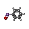

| #2: Chemical |  Mass: 96.063 Da / Num. of mol.: 2 / Source method: obtained synthetically / Formula: SO4 Mass: 96.063 Da / Num. of mol.: 2 / Source method: obtained synthetically / Formula: SO4#3: Chemical | ChemComp-CA / |  Mass: 40.078 Da / Num. of mol.: 1 / Source method: obtained synthetically / Formula: Ca Mass: 40.078 Da / Num. of mol.: 1 / Source method: obtained synthetically / Formula: Ca#4: Chemical |  Mass: 106.120 Da / Num. of mol.: 2 / Source method: obtained synthetically / Formula: C4H10O3 Mass: 106.120 Da / Num. of mol.: 2 / Source method: obtained synthetically / Formula: C4H10O3#5: Chemical | ChemComp-PA0 / |  Mass: 168.025 Da / Num. of mol.: 1 / Source method: obtained synthetically / Formula: C6H5AsO / Feature type: SUBJECT OF INVESTIGATION Mass: 168.025 Da / Num. of mol.: 1 / Source method: obtained synthetically / Formula: C6H5AsO / Feature type: SUBJECT OF INVESTIGATION#6: Water | ChemComp-HOH / | Mass: 18.015 Da / Num. of mol.: 250 / Source method: isolated from a natural source / Formula: H2O |
|---|
-Details
| Has ligand of interest | Y |
|---|---|
| Has protein modification | Y |
-Experimental details
-Experiment
| Experiment | Method: X-RAY DIFFRACTION / Number of used crystals: 1 |
|---|
- Sample preparation
| Crystal | Density Matthews: 3.13 Å3/Da / Density % sol: 60.8 % / Description: Cubic |
|---|---|
| Crystal grow | Temperature: 298 K / Method: vapor diffusion, sitting drop / pH: 8 / Details: 0.1M Tris, pH 8 0.2M Lithium sulfate 32% PEG3350 |
-Data collection
| Diffraction | Mean temperature: 100 K / Ambient temp details: NITROGEN STREAM / Serial crystal experiment: N |
|---|---|
| Diffraction source | Source: ROTATING ANODE / Type: RIGAKU FR-E SUPERBRIGHT / Wavelength: 1.5418 Å |
| Detector | Type: RIGAKU RAXIS HTC / Detector: IMAGE PLATE / Date: Jul 7, 2021 |
| Radiation | Protocol: SINGLE WAVELENGTH / Monochromatic (M) / Laue (L): M / Scattering type: x-ray |
| Radiation wavelength | Wavelength: 1.5418 Å / Relative weight: 1 |
| Reflection | Resolution: 1.81→33.9 Å / Num. obs: 30947 / % possible obs: 99.71 % / Redundancy: 17.5 % / Biso Wilson estimate: 23.4 Å2 / CC1/2: 0.999 / CC star: 1 / Rmerge(I) obs: 0.1114 / Rpim(I) all: 0.02724 / Rrim(I) all: 0.1148 / Net I/σ(I): 33.07 |
| Reflection shell | Resolution: 1.81→1.875 Å / Redundancy: 17.4 % / Rmerge(I) obs: 1.018 / Mean I/σ(I) obs: 3.8 / Num. unique obs: 3068 / CC1/2: 0.85 / CC star: 0.959 / Rpim(I) all: 0.25 / Rrim(I) all: 1.049 / % possible all: 100 |
- Processing
| Software |
| |||||||||||||||||||||||||||||||||||||||||||||||||||||||||||||||||||||||||||||||||||||||||||||||||||||||||
|---|---|---|---|---|---|---|---|---|---|---|---|---|---|---|---|---|---|---|---|---|---|---|---|---|---|---|---|---|---|---|---|---|---|---|---|---|---|---|---|---|---|---|---|---|---|---|---|---|---|---|---|---|---|---|---|---|---|---|---|---|---|---|---|---|---|---|---|---|---|---|---|---|---|---|---|---|---|---|---|---|---|---|---|---|---|---|---|---|---|---|---|---|---|---|---|---|---|---|---|---|---|---|---|---|---|---|
| Refinement | Method to determine structure: MOLECULAR REPLACEMENT Starting model: 1JC0 Resolution: 1.81→33.89 Å / SU ML: 0.1819 / Cross valid method: FREE R-VALUE / σ(F): 1.33 / Phase error: 19.8774 Stereochemistry target values: GeoStd + Monomer Library + CDL v1.2
| |||||||||||||||||||||||||||||||||||||||||||||||||||||||||||||||||||||||||||||||||||||||||||||||||||||||||
| Solvent computation | Shrinkage radii: 0.9 Å / VDW probe radii: 1.11 Å / Solvent model: FLAT BULK SOLVENT MODEL | |||||||||||||||||||||||||||||||||||||||||||||||||||||||||||||||||||||||||||||||||||||||||||||||||||||||||
| Displacement parameters | Biso mean: 28.23 Å2 | |||||||||||||||||||||||||||||||||||||||||||||||||||||||||||||||||||||||||||||||||||||||||||||||||||||||||
| Refinement step | Cycle: LAST / Resolution: 1.81→33.89 Å
| |||||||||||||||||||||||||||||||||||||||||||||||||||||||||||||||||||||||||||||||||||||||||||||||||||||||||
| Refine LS restraints |
| |||||||||||||||||||||||||||||||||||||||||||||||||||||||||||||||||||||||||||||||||||||||||||||||||||||||||
| LS refinement shell |
|
