 Movie
Movie Controller


+ Open data

- Basic information
| Entry | Database: PDB / ID: 8dsj | ||||||
|---|---|---|---|---|---|---|---|
| Title | Peptidylglycine alpha hydroxylating monooxygenase anaerobic | ||||||
 Components Components | Peptidylglycine alpha-amidating monooxygenase | ||||||
 Keywords Keywords | OXIDOREDUCTASE / copper / monooxygenase / peptidylglycine alpha hydroxylating | ||||||
| Function / homology |  Function and homology information Function and homology informationpeptidylglycine monooxygenase / peptidylamidoglycolate lyase / peptide amidation / peptidylglycine monooxygenase activity / peptidylamidoglycolate lyase activity / fatty acid primary amide biosynthetic process / ovulation cycle process / toxin metabolic process / long-chain fatty acid metabolic process / peptide metabolic process ...peptidylglycine monooxygenase / peptidylamidoglycolate lyase / peptide amidation / peptidylglycine monooxygenase activity / peptidylamidoglycolate lyase activity / fatty acid primary amide biosynthetic process / ovulation cycle process / toxin metabolic process / long-chain fatty acid metabolic process / peptide metabolic process / mitotic chromosome condensation / response to pH / L-ascorbic acid binding / limb development / response to zinc ion / response to copper ion / transport vesicle membrane / maternal process involved in female pregnancy / condensed chromosome / lactation / response to glucocorticoid / secretory granule / regulation of actin cytoskeleton organization / trans-Golgi network / response to estradiol / heart development / perikaryon / response to hypoxia / response to xenobiotic stimulus / copper ion binding / neuronal cell body / calcium ion binding / chromatin binding / regulation of transcription by RNA polymerase II / protein kinase binding / chromatin / perinuclear region of cytoplasm / cell surface / extracellular space / extracellular region / zinc ion binding / identical protein binding Similarity search - Function | ||||||
| Biological species |  | ||||||
| Method |  X-RAY DIFFRACTION / SYNCHROTRON / MOLECULAR REPLACEMENT / Resolution: 2.8 Å X-RAY DIFFRACTION / SYNCHROTRON / MOLECULAR REPLACEMENT / Resolution: 2.8 Å | ||||||
 Authors Authors | Arias, R.J. / Blackburn, N.J. | ||||||
| Funding support |  United States, 1items United States, 1items
| ||||||
 Citation Citation | Journal: Protein Sci. / Year: 2023 Title: New structures reveal flexible dynamics between the subdomains of peptidylglycine monooxygenase. Implications for an open to closed mechanism. Authors: Arias, R.J. / Welch, E.F. / Blackburn, N.J. | ||||||
| History |
|
- Structure visualization
| Structure viewer | Molecule: MolmilJmol/JSmol |
|---|
- Downloads & links
-Download
| PDBx/mmCIF format | 8dsj.cif.gz | 134.4 KB | Display | PDBx/mmCIF format |
|---|---|---|---|---|
| PDB format | pdb8dsj.ent.gz | 103.7 KB | Display | PDB format |
| PDBx/mmJSON format | 8dsj.json.gz | Tree view | PDBx/mmJSON format | |
| Others |  Other downloads Other downloads |
-Validation report
| Summary document | 8dsj_validation.pdf.gz | 2.4 MB | Display | wwPDB validaton report |
|---|---|---|---|---|
| Full document | 8dsj_full_validation.pdf.gz | 2.4 MB | Display | |
| Data in XML | 8dsj_validation.xml.gz | 26.9 KB | Display | |
| Data in CIF | 8dsj_validation.cif.gz | 33.9 KB | Display | |
| Arichive directory | https://data.pdbj.org/pub/pdb/validation_reports/ds/8dsjftp://data.pdbj.org/pub/pdb/validation_reports/ds/8dsj | HTTPS FTP |
-Related structure data
| Related structure data | 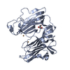 8dslC 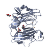 8dsnC 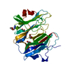 1phmS S: Starting model for refinement C: citing same article ( |
|---|---|
| Similar structure data |
-Links
 PDBj
PDBj


- Assembly
| Deposited unit | 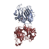
| ||||||||
|---|---|---|---|---|---|---|---|---|---|
| 1 | 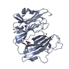
| ||||||||
| 2 | 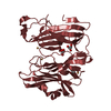
| ||||||||
| Unit cell |
|
-Components
| #1: Protein | Mass: 34577.641 Da / Num. of mol.: 2 Source method: isolated from a genetically manipulated source Source: (gene. exp.)  Cricetulus griseus (Chinese hamster) Cricetulus griseus (Chinese hamster)References: UniProt: P14925, peptidylglycine monooxygenase, peptidylamidoglycolate lyase #2: Chemical | ChemComp-CU /   Mass: 63.546 Da / Num. of mol.: 6 / Source method: obtained synthetically / Formula: Cu / Feature type: SUBJECT OF INVESTIGATION Mass: 63.546 Da / Num. of mol.: 6 / Source method: obtained synthetically / Formula: Cu / Feature type: SUBJECT OF INVESTIGATION#3: Chemical | ChemComp-GOL /   Mass: 92.094 Da / Num. of mol.: 5 / Source method: obtained synthetically / Formula: C3H8O3 Mass: 92.094 Da / Num. of mol.: 5 / Source method: obtained synthetically / Formula: C3H8O3Has ligand of interest | Y | Has protein modification | Y | |
|---|
-Experimental details
-Experiment
| Experiment | Method: X-RAY DIFFRACTION / Number of used crystals: 1 |
|---|
- Sample preparation
| Crystal | Density Matthews: 2.51 Å3/Da / Density % sol: 51.03 % |
|---|---|
| Crystal grow | Temperature: 298 K / Method: vapor diffusion, sitting drop / pH: 4.5 Details: 1.5 uL of WT PHM protein at 17 mg/mL in 20 mM sodium phosphate, pH 7.5 was added to 1.5 uL of mother liquor solution containing 16-18% PEG 20K, 20-250 mM sodium citrate, and 2 mM CuSO4. ...Details: 1.5 uL of WT PHM protein at 17 mg/mL in 20 mM sodium phosphate, pH 7.5 was added to 1.5 uL of mother liquor solution containing 16-18% PEG 20K, 20-250 mM sodium citrate, and 2 mM CuSO4. Plates were sealed using transparent tape. Crystals were formed within one week, and these initial crystals were used to seed succeeding trays using the same crystal conditions. Seeding was performed using a Hampton Research seed bead and Hampton Research seeding tool. Initial crystals (5-7 crystals) were vortexed with seed beads for 30 seconds in 30 uL mother liquor, and streaked into a new drop using the seeding tool. PH range: 4-5 |
-Data collection
| Diffraction | Mean temperature: 100 K / Serial crystal experiment: N | ||||||||||||||||||||||||
|---|---|---|---|---|---|---|---|---|---|---|---|---|---|---|---|---|---|---|---|---|---|---|---|---|---|
| Diffraction source | Source: SYNCHROTRON / Site: SSRL / Beamline: BL12-2 / Wavelength: 1 Å | ||||||||||||||||||||||||
| Detector | Type: DECTRIS PILATUS 6M / Detector: PIXEL / Date: Oct 27, 2021 | ||||||||||||||||||||||||
| Radiation | Protocol: SINGLE WAVELENGTH / Monochromatic (M) / Laue (L): M / Scattering type: x-ray | ||||||||||||||||||||||||
| Radiation wavelength | Wavelength: 1 Å / Relative weight: 1 | ||||||||||||||||||||||||
| Reflection | Resolution: 1.8→37.4 Å / Num. obs: 57073 / % possible obs: 92.8 % / Redundancy: 7.9 % / CC1/2: 0.855 / Rmerge(I) obs: 0.169 / Net I/σ(I): 3.6 | ||||||||||||||||||||||||
| Reflection shell | Diffraction-ID: 1
|
- Processing
| Software |
| ||||||||||||||||||||||||||||||||||||||||||||||||||||||||||||
|---|---|---|---|---|---|---|---|---|---|---|---|---|---|---|---|---|---|---|---|---|---|---|---|---|---|---|---|---|---|---|---|---|---|---|---|---|---|---|---|---|---|---|---|---|---|---|---|---|---|---|---|---|---|---|---|---|---|---|---|---|---|
| Refinement | Method to determine structure: MOLECULAR REPLACEMENT Starting model: 1phm Resolution: 2.8→37.4 Å / Cor.coef. Fo:Fc: 0.92 / Cor.coef. Fo:Fc free: 0.839 / SU B: 16.595 / SU ML: 0.333 / Cross valid method: THROUGHOUT / σ(F): 0 / ESU R Free: 0.437 / Stereochemistry target values: MAXIMUM LIKELIHOOD Details: HYDROGENS HAVE BEEN ADDED IN THE RIDING POSITIONS U VALUES : REFINED INDIVIDUALLY
| ||||||||||||||||||||||||||||||||||||||||||||||||||||||||||||
| Solvent computation | Ion probe radii: 0.8 Å / Shrinkage radii: 0.8 Å / VDW probe radii: 1.2 Å / Solvent model: MASK | ||||||||||||||||||||||||||||||||||||||||||||||||||||||||||||
| Displacement parameters | Biso max: 131.81 Å2 / Biso mean: 36.841 Å2 / Biso min: 9.61 Å2
| ||||||||||||||||||||||||||||||||||||||||||||||||||||||||||||
| Refinement step | Cycle: final / Resolution: 2.8→37.4 Å
| ||||||||||||||||||||||||||||||||||||||||||||||||||||||||||||
| Refine LS restraints |
| ||||||||||||||||||||||||||||||||||||||||||||||||||||||||||||
| LS refinement shell | Resolution: 2.8→2.872 Å / Rfactor Rfree error: 0 / Total num. of bins used: 20
|
